Japanese
English
- 有料閲覧
- Abstract 文献概要
- 1ページ目 Look Inside
I.はじめに
脳腫瘍の予後が,一般にその組織学的悪性度と相関することは周知の事実である.ところが1979年Kepesらは若年者に好発するgliomaのうち病理組織学的に腫瘍細胞のpleomorphismが著明で,一見悪性像を呈するが,良性のbiological behaviorを示す腫瘍があることに注目し,新しい腫瘍概念pleomorphic xanthoastrocytomaとして報告した4).本腫瘍の臨床および病理学上の意義は極めて大きいと思われるのにもかかわらず,いまだ本邦において報告されていない,最近われわれは,Kepesらと同様の症例を経験したので報告する.
A case of pleomorphic xanthoastrocytoma, the first case in Japan, is reported.
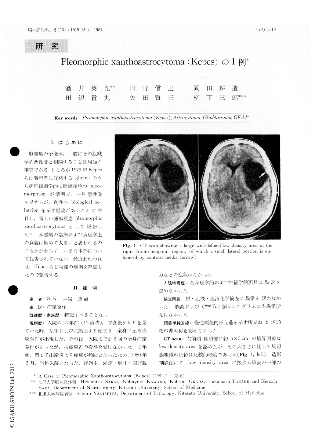
This is a 25-year-old woman with a history of convulsive seizures which were initiated on her left arm 13 years prior to admission. On admission, physical and neurological examinations revealed no abnormalities. CT-scan disclosed a large well-defined low density area in the right fronto-temporal region. A small peripheral portion of the low density area was enhanced by contrast media. The high density area located immediately beneath the inner table of the skull. Right carotid angiogram showed a large avascular area corresponding to the cystic lesion.

Copyright © 1981, Igaku-Shoin Ltd. All rights reserved.


