Japanese
English
研究
高血圧性脳内出血に関する研究(第2報)—血腫の経時的追跡およびCT分類について
A Study of Hypertensive Intracerehral Hemorrhage (Ⅱ) : Sequential CT Examination and Classification
三浦 直久
1
,
中原 明
1
,
加川 瑞夫
1
,
喜多村 孝一
1
,
小林 直紀
2
Naohisa MIURA
1
,
Akira NAKAHARA
1
,
Mizuo KAGAWA
1
,
Koichi KITAMURA
1
,
Naotoshi KOBAYASHI
2
1東京女医科大学脳神経センター脳神経外科
2東京女医科大学脳神経センター神経放射線科
1Department of Neurosurgery, Institute of Neurology, Tokyo Women's Medical College
2Department of Neuroradiology, Institute of Neurology, Tokyo Women's Medical College
キーワード:
Computerized tomography
,
EMI scan
,
Hypertensive intracerebral hemorrhage
Keyword:
Computerized tomography
,
EMI scan
,
Hypertensive intracerebral hemorrhage
pp.635-645
発行日 1978年7月10日
Published Date 1978/7/10
DOI https://doi.org/10.11477/mf.1436200836
- 有料閲覧
PPV購入案内
1,320円 (1,200円+税10%) こちらは、635ページから645ページの文献です。
PPV(ペイ・パー・ビュー)とは、論文(記事)単位で購入・閲覧ができるサービスです。
購入には医書.jp本会員登録が必要です。
会員登録完了後に改めて上記「購入サイトへ」を選択してください。
または「購入サイトへ」選択後に「購入ログイン」、「新規会員登録」の順に進んでください。
会員登録について詳しくはをご確認ください。
購入には医書.jp本会員登録が必要です。
会員登録完了後に改めて上記「購入サイトへ」を選択してください。
または「購入サイトへ」選択後に「購入ログイン」、「新規会員登録」の順に進んでください。
会員登録について詳しくはをご確認ください。
- Abstract 文献概要
- 1ページ目 Look Inside
Ⅰ.はじめに
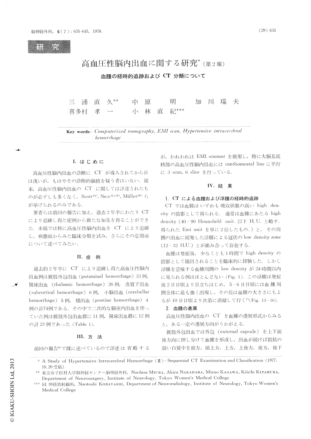
高血圧性脳内出血の診断にCTが導人されてから日は浅いが,もはやその診断的価値を疑う者はいない.従来,高血圧性脳内出血のCTに関しては詳述されたものが必ずしも多くなく,Scott14),New12,13),Muller10)らが挙げられるのみである.
著者らは前回の報告に加え,過去2年半にわたりCTにより追跡し得た症例から新たな知見を得ることができた.本稿では特に高血圧性脳内出血をCTにより追跡し,病態面からみた臨床分類を試み,さらにその応用面について述べてみたい.
We experienced 74 patients with hypertensive intracerebral and posterior fossa hemorrhage during last 30 months.
1) Two to three days after hemorrhage, a low density zone was seen at the anterior and posterior pole of the high density area. About 5 days after hemorrhage, high density area was present with wide surrounding zone of edema and then gradually decreased in size of the high density area with a wide zone of surrounding low density of liquefied hematoma and/or edema over 15-days period.
Copyright © 1978, Igaku-Shoin Ltd. All rights reserved.