Japanese
English
- 有料閲覧
PPV購入案内
1,320円 (1,200円+税10%) こちらは、1091ページから1097ページの文献です。購入には医書.jp本会員登録が必要です。
会員登録完了後に改めて上記「購入サイトへ」を選択してください。
または「購入サイトへ」選択後に「購入ログイン」、「新規会員登録」の順に進んでください。
会員登録について詳しくはをご確認ください。
- Abstract 文献概要
- 1ページ目 Look Inside
- 参考文献 Reference
Ⅰ.はじめに
黄色肉芽腫(xanthogranuloma)は1900年にBlumerによって初めて報告された4).コレステロール裂,マクロファージ(xanthoma cells),ヘモジデリン沈着物などにおける慢性炎症の細胞性反応を示し,細胞質内に脂質(lipid)の蓄積を呈する多様な性質をもつとされる良性腫瘤である.変性,炎症性,代謝性,腫瘍性など多様な性質が認められ,脳出血,脳梗塞,感染,壊死などから二次性に発生することが多いとされている.頭蓋内では脈絡叢から発生することが多く,側脳室7),第三脳室12)に認められる報告が多く,その他に鞍上部2,16),松果体部19)での報告,下垂体腺腫でも黄色肉芽腫様変化に富む例13)が散見される.脈絡叢を除く脳内の報告は少ない5,9,15).
今回,われわれは特異的な画像所見を呈した前頭葉腫瘤で,病理組織学的に黄色肉芽腫であった1例を経験し,その画像特徴を含め考察したので報告する.
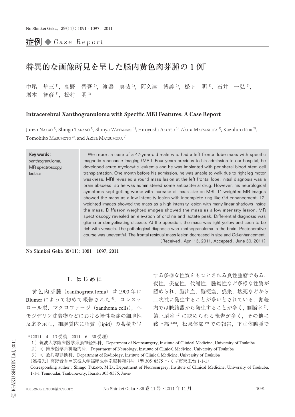
We report a case of a 47-year-old male who had a left frontal lobe mass with specific magnetic resonance imaging (MRI). Four years previous to his admission to our hospital,he developed acute myelocytic leukemia and he was implanted with peripheral blood stem cell transplantation. One month before his admission,he was unable to walk due to right leg motor weakness. MRI revealed a round mass lesion at the left frontal lobe. Initial diagnosis was a brain abscess,so he was administered some antibacterial drug. However,his neurological symptoms kept getting worse with increase of mass size on MRI. T1-weighted MR images showed the mass as a low intensity lesion with incomplete ring-like Gd-enhancement. T2-weighted images showed the mass as a high intensity lesion with many linear shadows inside the mass. Diffusion weighted images showed the mass as a low intensity lesion. MR spectroscopy revealed an elevation of choline and lactate peak. Differential diagnosis was glioma or demyelinating disease. At the operation,the mass was light yellow and seen to be rich with vessels. The pathological diagnosis was xanthogranuloma in the brain. Postoperative course was uneventful. The frontal residual mass lesion decreased in size and Gd-enhancement.
Copyright © 2011, Igaku-Shoin Ltd. All rights reserved.