Japanese
English
- 有料閲覧
PPV購入案内
1,320円 (1,200円+税10%) こちらは、697ページから702ページの文献です。購入には医書.jp本会員登録が必要です。
会員登録完了後に改めて上記「購入サイトへ」を選択してください。
または「購入サイトへ」選択後に「購入ログイン」、「新規会員登録」の順に進んでください。
会員登録について詳しくはをご確認ください。
- Abstract 文献概要
- 1ページ目 Look Inside
- 参考文献 Reference
Ⅰ.はじめに
Solitary fibrous tumor(SFT)は主に胸膜発生で知られる間葉系細胞由来の腫瘍性病変であり,1931年にKlempererとRabinにより初めて報告された8).全身のあらゆる臓器に発生することが明らかとなってきたが,中枢神経系における最初の報告は,1996年のCarneiroらが髄膜起源とした例である1).現在まで脊髄を含め中枢神経系発生症例の報告が散見され,臨床上はmeningiomaやhemanigopericytomaとの鑑別が重要とされている3,16,19).2000年のWHO分類では髄膜発生腫瘍として新たに分類され17),その発生母地を硬膜とする報告も多いが2,3),頭蓋内腫瘍性病変としては依然として稀な腫瘍であり,いまだ確立されたものではない.今回われわれは,その画像と術中所見が発生母地を考える上で興味深い所見を示した円蓋部SFTの1例を経験したので,過去の文献をふまえて報告する.
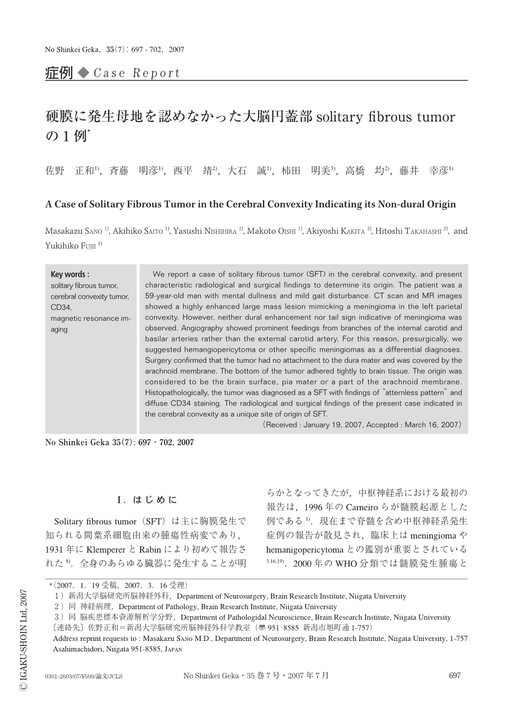
We report a case of solitary fibrous tumor (SFT) in the cerebral convexity, and present characteristic radiological and surgical findings to determine its origin. The patient was a 59-year-old man with mental dullness and mild gait disturbance. CT scan and MR images showed a highly enhanced large mass lesion mimicking a meningioma in the left parietal convexity. However, neither dural enhancement nor tail sign indicative of meningioma was observed. Angiography showed prominent feedings from branches of the internal carotid and basilar arteries rather than the external carotid artery. For this reason, presurgically, we suggested hemangiopericytoma or other specific meningiomas as a differential diagnoses. Surgery confirmed that the tumor had no attachment to the dura mater and was covered by the arachnoid membrane. The bottom of the tumor adhered tightly to brain tissue. The origin was considered to be the brain surface, pia mater or a part of the arachnoid membrane. Histopathologically, the tumor was diagnosed as a SFT with findings of “atternless pattern” and diffuse CD34 staining. The radiological and surgical findings of the present case indicated in the cerebral convexity as a unique site of origin of SFT.
Copyright © 2007, Igaku-Shoin Ltd. All rights reserved.