Japanese
English
- 有料閲覧
- Abstract 文献概要
- 1ページ目 Look Inside
- 参考文献 Reference
Ⅰ.はじめに
Solitary fibrous tumor(SFT)は,1931年に胸膜原発の症例が報告されたのが最初である.当初は胸膜起源のものと理解されていたが,その後同様の組織像をもつ腫瘍が胸膜以外にも報告されるようになった.組織像が似ていることでhemangiopericytomaとの同異が問題となることがある.WHO軟部組織腫瘍分類では同一の疾患概念として記載されているが,WHO脳腫瘍分類ではそれぞれ別の疾患に分類されており,SFTの疾患概念の確立は今後の課題であると考えられる.
中枢神経系に発生するSFTは現在まで200例以上の報告があるが,トルコ鞍内発生SFTは,われわれが渉猟し得た範囲では4例の報告があるのみであり,極めて稀な症例である.これらは非機能性下垂体腺腫と診断されることが多いが,治療戦略上その鑑別は重要である.トルコ鞍内発生SFTの画像所見には特徴があり,典型的な症例では正確な術前診断も可能であると考える.
今回われわれはトルコ鞍内を発生母地とし,非機能性下垂体腺腫との鑑別を要した1例を経験したので,非機能性下垂体腺腫との鑑別点を中心に文献的考察を踏まえて報告する.
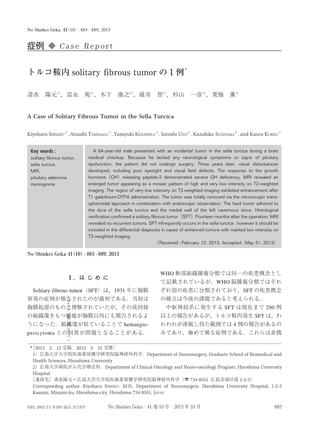
A 64-year-old male presented with an incidental tumor in the sella turcica during a brain medical checkup. Because he lacked any neurological symptoms or signs of pituitary dysfunction, the patient did not undergo surgery. Three years later, visual disturbances developed, including poor eyesight and visual field defects. The response to the growth hormone (GH) releasing peptide-2 demonstrated severe GH deficiency. MRI revealed an enlarged tumor appearing as a mosaic pattern of high and very low intensity on T2-weighted imaging. The region of very low intensity on T2-weighted imaging exhibited enhancement after T1 gadolinium-DTPA administration. The tumor was totally removed via the microscopic trans-sphenoidal approach in combination with endoscopic observation. The hard tumor adhered to the dura of the sella turcica and the medial wall of the left cavernous sinus. Histological verification confirmed a solitary fibrous tumor (SFT). Fourteen months after the operation, MRI revealed no recurrent tumors. SFT infrequently occurs in the sella turcica;however it should be included in the differential diagnosis in cases of enhanced tumors with marked low intensity on T2-weighted imaging.

Copyright © 2013, Igaku-Shoin Ltd. All rights reserved.


