Japanese
English
- 有料閲覧
PPV購入案内
1,320円 (1,200円+税10%) こちらは、486ページから494ページの文献です。購入には医書.jp本会員登録が必要です。
会員登録完了後に改めて上記「購入サイトへ」を選択してください。
または「購入サイトへ」選択後に「購入ログイン」、「新規会員登録」の順に進んでください。
会員登録について詳しくはをご確認ください。
- Abstract 文献概要
- 1ページ目 Look Inside
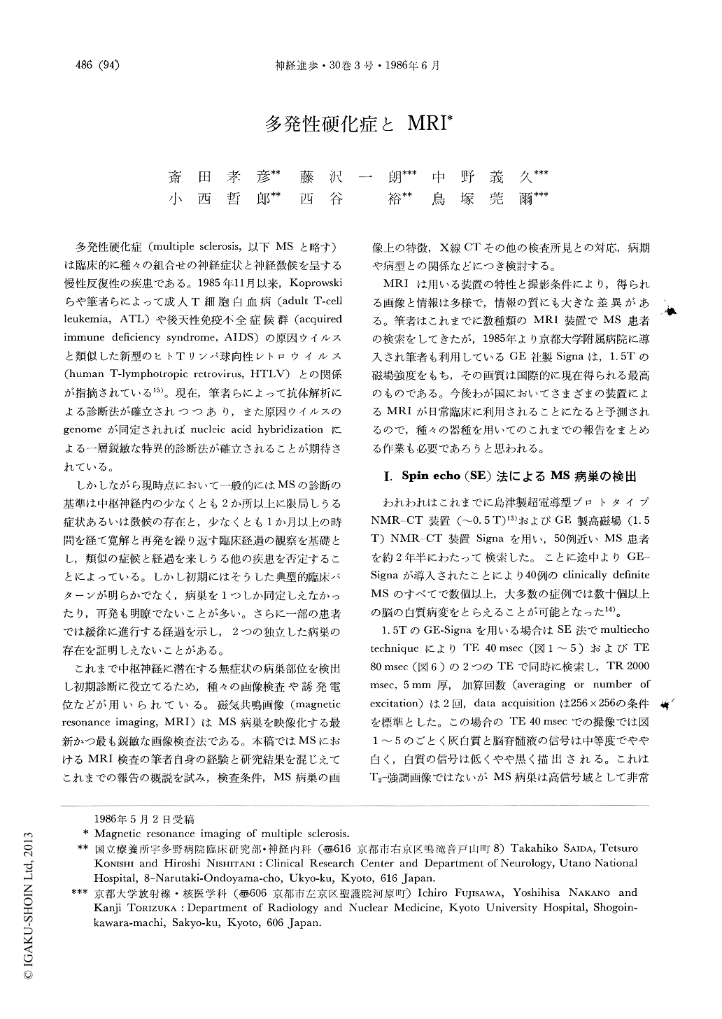
多発性硬化症(multiple sclerosis,以下MSと略す)は臨床的に種々の組合せの神経症状と神経徴候を呈する慢性反復性の疾患である。1985年11月以来,Koprowskiらや筆者らによって成人T細胞白血病(adult T-cell leukemia,ATL)や後天性免疫不全症候群(acquired immune deficiency syndrome,AIDS)の原因ウイルスと類似した新型のヒトTリンパ球向性レトロウイルス(human T-lymphotropic retrovirus,HTLV)との関係が指摘されている15)。現在,筆者らによって抗体解析による診断法が確立されつつあり,また原因ウイルスのgenomeが同定されればnucleic acid hybridizationによる一層鋭敏な特異的診断法が確立されることが期待されている。
しかしながら現時点において一般的にはMSの診断の基準は中枢神経内の少なくとも2か所以上に限局しうる症状あるいは徴候の存在と,少なくとも1か月以上の時間を経て寛解と再発を繰り返す臨床経過の観察を基礎とし,類似の症候と経過を来しうる他の疾患を否定することによっている。しかし初期にはそうした典型的臨床パターンが明らかでなく,病巣を1つしか同定しえなかったり,再発も明瞭でないことが多い。さらに一部の患者では緩徐に進行する経過を示し,2つの独立した病巣の存在を証明しえないことがある。
Forty clinically definite multiple sclerosis (MS) patients and 10 possible MS patients were studied during last 2 years with a 0.5 tesla superconductive Shimazu prototype MRI scanner and later with a 1.5 tesla high resolution MRI scanner, GE-Signa. All clinically definite MS patients showed multiple white matter lesions which are usually best visualized by the spin-echo technique. Most MRI detected cerebral lesions were asymptomatic and the patients with spinal and opticospinal type MS, which are relatively more frequent among Japanese MS, also had a number of subclinical cerebral lesions. It appears likely that MRI detected lesions usually do not resolve completely and consequently changes seen with this neuroimaging technique may represent the cumulative effects of past and present disease activity.
Copyright © 1986, Igaku-Shoin Ltd. All rights reserved.