Japanese
English
- 有料閲覧
- Abstract 文献概要
- 1ページ目 Look Inside
はじめに
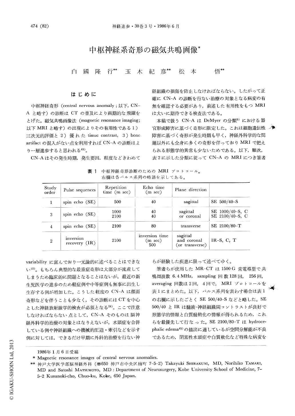
中枢神経奇形(central nervous anomaly;以下,CN-Aと略す)の診断はCTの普及により画期的な飛躍をとげた。磁気共鳴画像法(magnetic resonance imaging;以下MRIと略す)の出現によりその有用性である 1)三次元的評価と 2)優れたtissue contrast,3)boneartifactの混入がない点を利用すればCN-Aの診断はより一層進歩すると思われる43)。
CN-Aはその発生時期,発生要因,程度などきわめてvariabilityに富んでおり一元論的に述べることはできない12)。もちろん典型的な最重症奇形は大部分が流産してしまうため臨床的に問題となることはないが,最近の新生児医学の進歩のため軽症例や中等症例も無事に出生し生存する例が増加した。こうした軽度のCN-Aは顔面奇形などを伴うことも少なく,その診断にはCTを中心とした神経放射線学的検査が必須となる24)。ここで注意しなければならない点として,CN-Aそのものは脳神経外科学的治療の対象とはなりえないが,水頭症を合併している例や神経組織への機械的圧迫・牽引などを示す例に対しては,できるだけ早期に外科的治療を行ない神経組織の損傷を防止しなければならない。したがって正確にCN-Aの診断を行ない治療の対象となる病変の有無を確認する必要があり,前述した有用性をもつMRIは大いに期待できる検査法である。
Magnetic resonance (MR) imaging is useful technique to make a diagnosis of congenital anomalies of the central nervous system as well as other nervous diseases with excellent three-dimensional spatial resolution and without bone artifacts. We had experiences of MR diagnosis of various central nervous anomalies, demonstrated and described characteristic findings in each anomaly.
In Arnold-Chiari deformity, MR imaging revealed a morphological differences between type I and type II deformities; the latter had many supratentorial anomalies in addition to characteristic abnormalities in posterior fossa structures.

Copyright © 1986, Igaku-Shoin Ltd. All rights reserved.


