Japanese
English
- 有料閲覧
- Abstract 文献概要
- 1ページ目 Look Inside
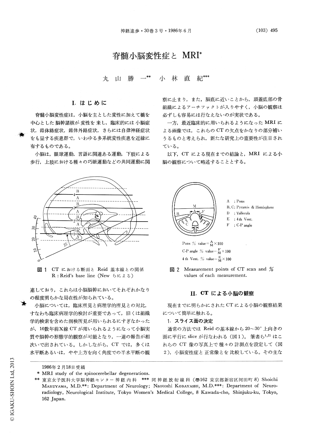
I.はじめに
脊髄小脳変性症は,小脳を主とした変性に加えて橋を中心とした脳幹諸核が変性を来し,臨床的には小脳症状,錐体路症状,錐体外路症状,さらには自律神経症状をも呈する疾患群で,いわゆる多系統変性疾患を近縁に有するものである。
小脳は,眼球運動,言語に関連ある運動,下肢による歩行,上肢における種々の巧緻運動などの共同運動に関連しており,これらは小脳脳幹においてそれぞれかなりの程度明らかな局在性が知られている。
Clinical cases of spinocerebellar degenerations were observed by Magnetic Resonance Imaging (MRI) method.
MRI is very useful for the observations of the cerebellum by changing the parameters of the imaging easily and we can chose the optimal conditions for them.
For the topographic observations of the lesions in the spinocerebellar degenerations, MRI is very helpful for three dimensional reconstruction because the saggital sections of the cerebellum can be taken easily. These section can identified the vermis, the anterior and lateral cerebellar lobes, nodules and basal lobes of which CTscan can not realize the exact structures. We can compare these pathoanatomical results with the clinical findings of the spinocerebellar degenerations and can analyzed the clinical and pathoanatomical correlations.
Copyright © 1986, Igaku-Shoin Ltd. All rights reserved.