Japanese
English
- 有料閲覧
PPV購入案内
1,320円 (1,200円+税10%) こちらは、17ページから25ページの文献です。購入には医書.jp本会員登録が必要です。
会員登録完了後に改めて上記「購入サイトへ」を選択してください。
または「購入サイトへ」選択後に「購入ログイン」、「新規会員登録」の順に進んでください。
会員登録について詳しくはをご確認ください。
- Abstract 文献概要
- 1ページ目 Look Inside
- 参考文献 Reference
はじめに
頭部に発生する先天性血管奇形は,古典的に脳動静脈奇形(arteriovenous malformation),毛細血管拡張症(capillary telangiectasia),海綿状血管腫(cavernous [hem]angioma, cavernous malformation, cavernoma),静脈性血管腫(venous angioma, venous malformation)の4つに分類される。これらのうち,本稿では海綿状血管腫,静脈性血管腫に関して,疫学,症状,一般的な治療法を中心に述べる。なお画像所見や定位的放射線治療の詳細,脳幹部海綿状血管腫の外科的治療に関する話題は本号他稿に譲る。
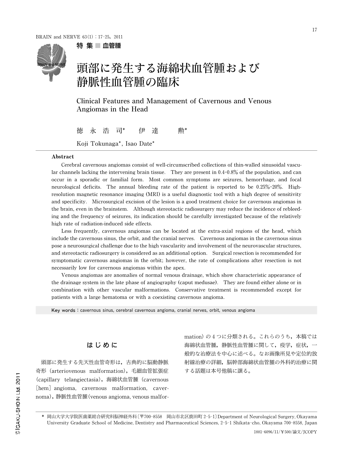
Abstract
Cerebral cavernous angiomas consist of well-circumscribed collections of thin-walled sinusoidal vascular channels lacking the intervening brain tissue. They are present in 0.4-0.8% of the population, and can occur in a sporadic or familial form. Most common symptoms are seizures, hemorrhage, and focal neurological deficits. The annual bleeding rate of the patient is reported to be 0.25%-20%. High-resolution magnetic resonance imaging (MRI) is a useful diagnostic tool with a high degree of sensitivity and specificity. Microsurgical excision of the lesion is a good treatment choice for cavernous angiomas in the brain, even in the brainstem. Although stereotactic radiosurgery may reduce the incidence of rebleeding and the frequency of seizures, its indication should be carefully investigated because of the relatively high rate of radiation-induced side effects.
Less frequently, cavernous angiomas can be located at the extra-axial regions of the head, which include the cavernous sinus, the orbit, and the cranial nerves. Cavernous angiomas in the cavernous sinus pose a neurosurgical challenge due to the high vascularity and involvement of the neurovascular structures, and stereotactic radiosurgery is considered as an additional option. Surgical resection is recommended for symptomatic cavernous angiomas in the orbit; however, the rate of complications after resection is not necessarily low for cavernous angiomas within the apex.
Venous angiomas are anomalies of normal venous drainage,which show characteristic appearance of the drainage system in the late phase of angiography (caput medusae). They are found either alone or in combination with other vascular malformations. Conservative treatment is recommended except for patients with a large hematoma or with a coexisting cavernous angioma.
Copyright © 2011, Igaku-Shoin Ltd. All rights reserved.