Japanese
English
- 有料閲覧
PPV購入案内
1,320円 (1,200円+税10%) こちらは、371ページから375ページの文献です。購入には医書.jp本会員登録が必要です。
会員登録完了後に改めて上記「購入サイトへ」を選択してください。
または「購入サイトへ」選択後に「購入ログイン」、「新規会員登録」の順に進んでください。
会員登録について詳しくはをご確認ください。
- Abstract 文献概要
- 1ページ目 Look Inside
I.はじめに
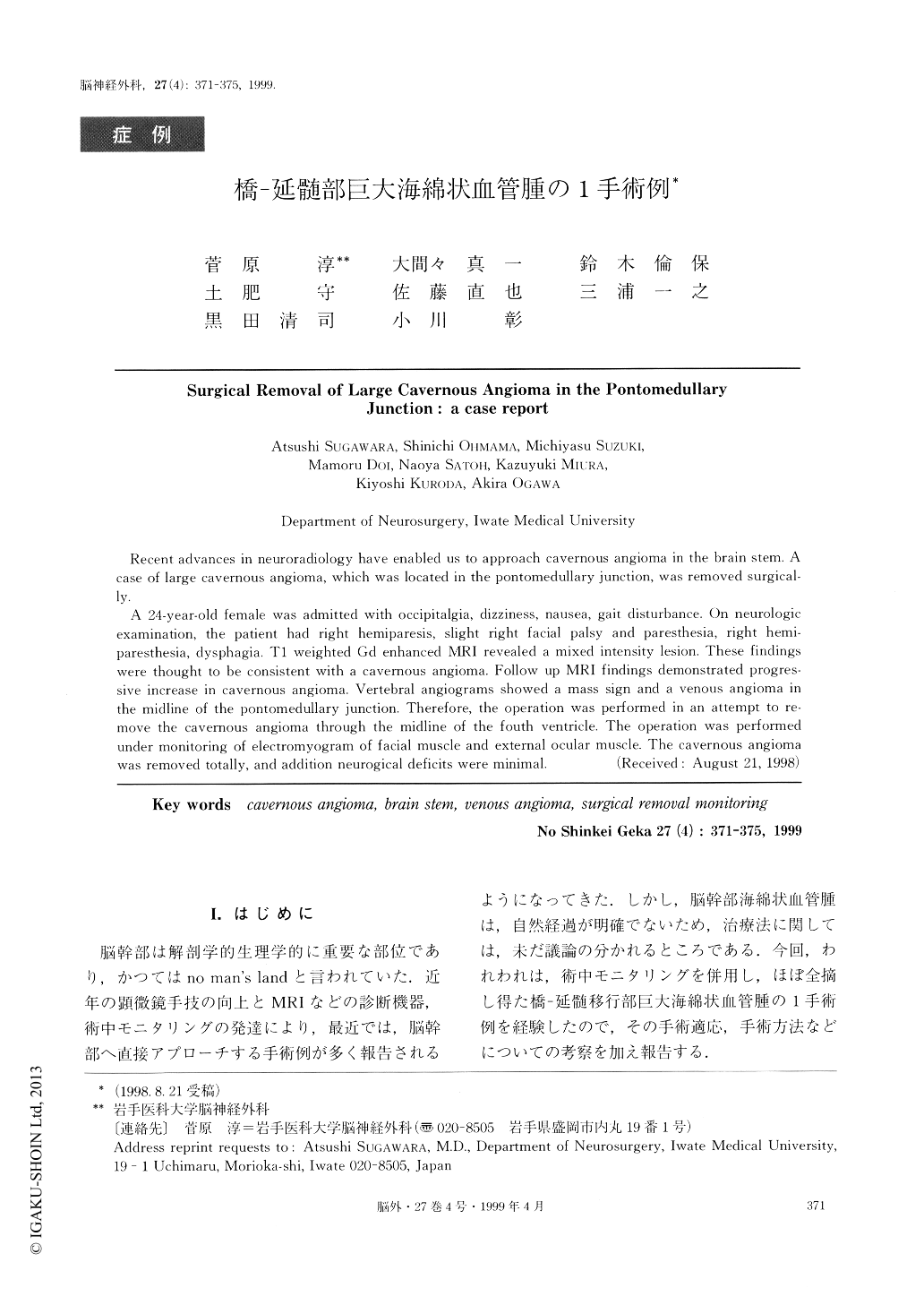
脳幹部は解剖学的生理学的に重要な部位であり,かつてはno man's landと言われていた.近年の顕微鏡手技の向上とMRIなどの診断機器,術中モニタリングの発達により,最近では,脳幹部へ直接アプローチする手術例が多く報告されるようになってきた.しかし,脳幹部海綿状血管腫は,自然経過が明確でないため,治療法に関しては,未だ議論の分かれるところである.今回,われわれは,術中モニタリングを併用し,ほぼ全摘し得た橋-延髄移行部巨大海綿状血管腫の1手術例を経験したので,その手術適応,手術方法などについての考察を加え報告する.
Recent advances in neuroradiology have enabled us to approach cavernous angioma in the brain stem. Acase of large cavernous angioma, which was located in the pontomedullary junction, was removed surgical-ly.
A 24-year-old female was admitted with occipitalgia, dizziness, nausea, gait disturbance. On neurologicexamination, the patient had right hemiparesis, slight right facial palsy and paresthesia, right hemi-paresthesia, dysphagia. T1 weighted Gd enhanced MRI revealed a mixed intensity lesion. These findingswere thought to be consistent with a cavernous angioma. Follow up MRI findings demonstrated progres-sive increase in cavernous angioma. Vertebral angiograms showed a mass sign and a venous angioma inthe midline of the pontomedullary junction. Therefore, the operation was performed in an attempt to re-move the cavernous angioma through the midline of the fouth ventricle. The operation was performedunder monitoring of electromyogram of facial muscle and external ocular muscle. The cavernous angiomawas removed totally, and addition neurogical deficits were minimal.
Copyright © 1999, Igaku-Shoin Ltd. All rights reserved.