Japanese
English
- 有料閲覧
PPV購入案内
1,320円 (1,200円+税10%) こちらは、245ページから249ページの文献です。購入には医書.jp本会員登録が必要です。
会員登録完了後に改めて上記「購入サイトへ」を選択してください。
または「購入サイトへ」選択後に「購入ログイン」、「新規会員登録」の順に進んでください。
会員登録について詳しくはをご確認ください。
- Abstract 文献概要
- 1ページ目 Look Inside
- 参考文献 Reference
Ⅰ.はじめに
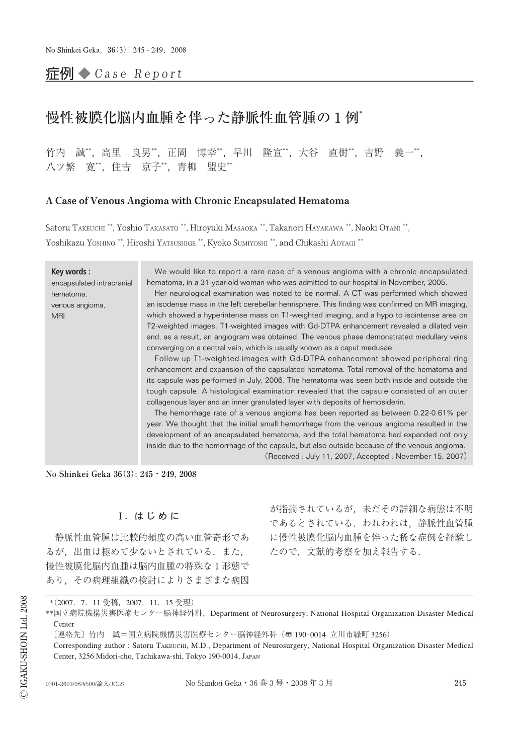
静脈性血管腫は比較的頻度の高い血管奇形であるが,出血は極めて少ないとされている.また,慢性被膜化脳内血腫は脳内血腫の特殊な1形態であり,その病理組織の検討によりさまざまな病因が指摘されているが,未だその詳細な病態は不明であるとされている.われわれは,静脈性血管腫に慢性被膜化脳内血腫を伴った稀な症例を経験したので,文献的考察を加え報告する.
We would like to report a rare case of a venous angioma with a chronic encapsulated hematoma, in a 31-year-old woman who was admitted to our hospital in November, 2005.
Her neurological examination was noted to be normal. A CT was performed which showed an isodense mass in the left cerebellar hemisphere. This finding was confirmed on MR imaging, which showed a hyperintense mass on T1-weighted imaging, and a hypo to isointense area on T2-weighted images. T1-weighted images with Gd-DTPA enhancement revealed a dilated vein and, as a result, an angiogram was obtained. The venous phase demonstrated medullary veins converging on a central vein, which is usually known as a caput medusae.
Follow up T1-weighted images with Gd-DTPA enhancement showed peripheral ring enhancement and expansion of the capsulated hematoma. Total removal of the hematoma and its capsule was performed in July, 2006. The hematoma was seen both inside and outside the tough capsule. A histological examination revealed that the capsule consisted of an outer collagenous layer and an inner granulated layer with deposits of hemosiderin.
The hemorrhage rate of a venous angioma has been reported as between 0.22-0.61% per year. We thought that the initial small hemorrhage from the venous angioma resulted in the development of an encapsulated hematoma, and the total hematoma had expanded not only inside due to the hemorrhage of the capsule, but also outside because of the venous angioma.
Copyright © 2008, Igaku-Shoin Ltd. All rights reserved.