Japanese
English
症例
腎部分切除術を施行した大豆大の腎細胞癌
PARTIAL NEPHRECTOMY OF A SMALL (7 MM) INCIDENTAL RENAL CELL CARCINOMA
高山 達也
1
,
伊原 博行
1
,
福田 健
1
,
畑 昌宏
1
,
鈴木 和雄
2
,
藤田 公生
2
Tatsuya Takayama
1
,
Hiroyuki Ihara
1
,
Ken Fukuta
1
,
Masahiro Hata
1
,
Kazuo Suzuki
2
,
Kimio Fujita
2
1聖隷三方原病院泌尿器科
2浜松医科大学泌尿器科
キーワード:
腎部分切除術
Keyword:
腎部分切除術
pp.1031-1034
発行日 1995年12月20日
Published Date 1995/12/20
DOI https://doi.org/10.11477/mf.1413901649
- 有料閲覧
PPV購入案内
1,320円 (1,200円+税10%) こちらは、1031ページから1034ページの文献です。
PPV(ペイ・パー・ビュー)とは、論文(記事)単位で購入・閲覧ができるサービスです。
購入には医書.jp本会員登録が必要です。
会員登録完了後に改めて上記「購入サイトへ」を選択してください。
または「購入サイトへ」選択後に「購入ログイン」、「新規会員登録」の順に進んでください。
会員登録について詳しくはをご確認ください。
購入には医書.jp本会員登録が必要です。
会員登録完了後に改めて上記「購入サイトへ」を選択してください。
または「購入サイトへ」選択後に「購入ログイン」、「新規会員登録」の順に進んでください。
会員登録について詳しくはをご確認ください。
- Abstract 文献概要
- 1ページ目 Look Inside
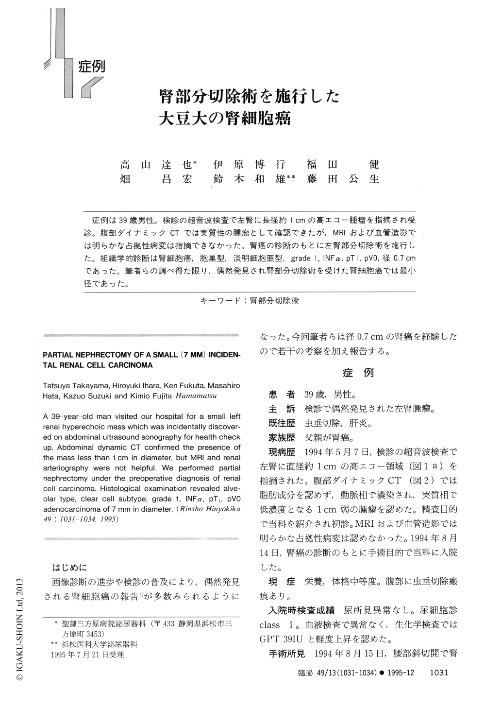
症例は39歳男性。検診の超音波検査で左腎に長径約1cmの高エコー腫瘤を指摘され受診。腹部ダイナミックCTでは実質性の腫瘤として確認できたが,MRIおよび血管造影では明らかな占拠性病変は指摘できなかった。腎癌の診断のもとに左腎部分切除術を施行した。組織学的診断は腎細胞癌,胞巣型,淡明細胞亜型,grade 1,INFα,pT 1,pVO,径0.7 cmであった。筆者らの調べ得た限り,偶然発見され腎部分切除術を受けた腎細胞癌では最小径であった。
A 39-year-old man visited our hospital for a small left renal hyperechoic mass which was incidentally discovered on abdominal ultrasound sonography for health check up. Abdominal dynamic CT confirmed the presence of the mass less than 1 cm in diameter, but MRI and renal arteriography were not helpful. We performed partial nephrectomy under the preoperative diagnosis of renal cell carcinoma. Histological examination revealed alveolar type, clear cell subtype, grade 1, INFa, pT1, pV0 adenocarcinoma of 7 mm in diameter.
Copyright © 1995, Igaku-Shoin Ltd. All rights reserved.