Japanese
English
- 有料閲覧
PPV購入案内
1,320円 (1,200円+税10%) こちらは、303ページから309ページの文献です。購入には医書.jp本会員登録が必要です。
会員登録完了後に改めて上記「購入サイトへ」を選択してください。
または「購入サイトへ」選択後に「購入ログイン」、「新規会員登録」の順に進んでください。
会員登録について詳しくはをご確認ください。
- Abstract 文献概要
- 1ページ目 Look Inside
はじめに
Iversen,Brun7)らが1951年に吸引法による経皮的腎生検法を発表したのち,肝生検に使用されていたSilvermann針がこれに応用され,更にMue-hrcke9),Kark17)らにより静脈性腎盂撮影像をもととして腎下極を作図上求める方法が確立されてから経皮的腎生検法は広く普及するようになつた。
とはいえMuehrckeらの経皮的腎生検法でも常に組織診断に十分な量の腎組織を確実かつ安全に採取しうるわけでなく欧米で50〜98%7,9,18,22),本邦でも40〜97%11,19,24)にも変動する成功率が報告されている。
また,副作用10,22)については強度の血尿,腎周囲血腫,腎茎部損傷,腎梗塞などによる腎摘除例,気胸,血胸,腎動静脈瘻,尿管損傷1),肝・脾・胆嚢・大腸などの損傷例,更には少数ではあるが死亡例12,22)すら報告されている。
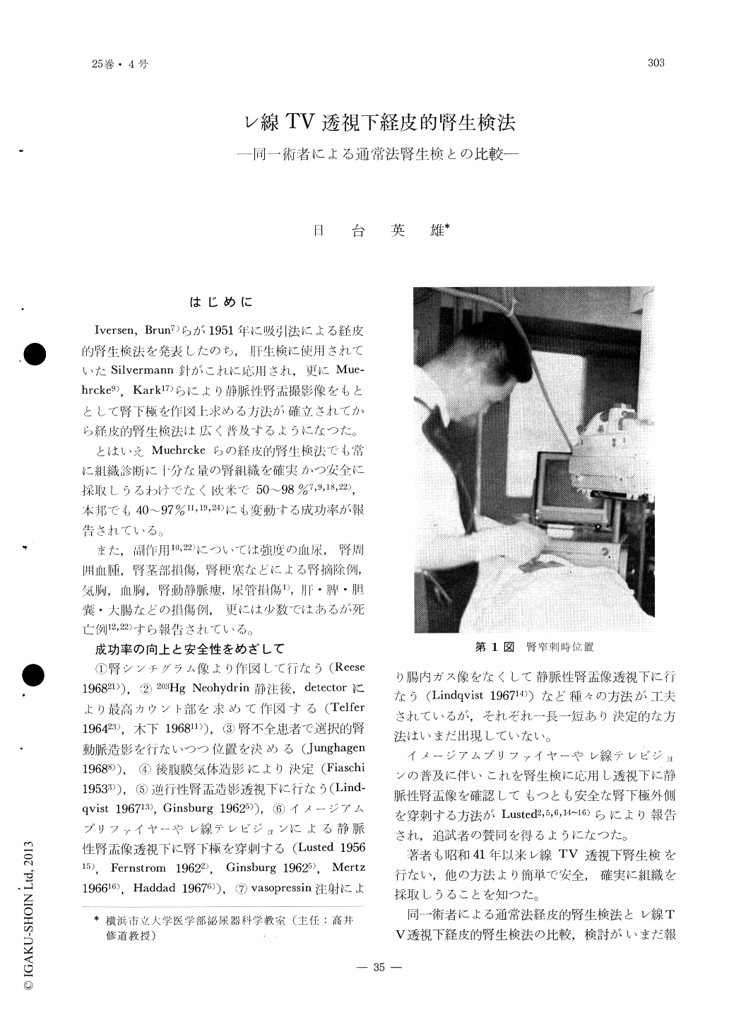
The author performed percutanous renal biopsy on 17 cases by the routine procedure (Kark's method) and another 17 cases under direct fluoroscopic control by roentgen television.
Under fluoroscopic control, renal tissues containing 20 glomeruli on the average were biopsied in all cases without severe sequela, whereas the routine method failed to obtain any renal tissue in 33%.
Comparison of two series disclosed, that enough renal tissue, adequate for histopathological diagnosis, can be obtained by the method under roentgen television fluoroscopy with accuracy and safety, by the same proficiency.
Copyright © 1971, Igaku-Shoin Ltd. All rights reserved.