Japanese
English
- 有料閲覧
PPV購入案内
1,320円 (1,200円+税10%) こちらは、979ページから982ページの文献です。購入には医書.jp本会員登録が必要です。
会員登録完了後に改めて上記「購入サイトへ」を選択してください。
または「購入サイトへ」選択後に「購入ログイン」、「新規会員登録」の順に進んでください。
会員登録について詳しくはをご確認ください。
- Abstract 文献概要
- 1ページ目 Look Inside
- 参考文献 Reference
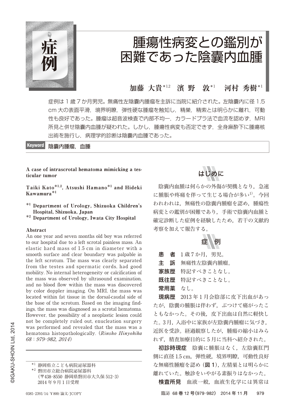
症例は1歳7か月男児。無痛性左陰囊内腫瘤を主訴に当院に紹介された。左陰囊内に径1.5cm大の表面平滑,境界明瞭,弾性硬な腫瘤を触知し,精巣,精索とは明らかに離れ,可動性も良好であった。腫瘤は超音波検査で内部不均一,カラードプラ法で血流を認めず,MRI所見と併せ陰囊内血腫が疑われた。しかし,腫瘍性病変も否定できず,全身麻酔下に腫瘍核出術を施行し,病理学的診断は陰囊内血腫であった。
An one year and seven months old boy was referred to our hospital due to a left scrotal painless mass. An elastic hard mass of 1.5 cm in diameter with a smooth surface and clear boundary was palpable in the left scrotum. The mass was clearly separated from the testes and spermatic cords, had good mobility. No internal heterogeneity or calcification of the mass was observed by ultrasound examination, and no blood flow within the mass was discovered by color doppler imaging. On MRI, the mass was located within fat tissue in the dorsal-caudal side of the base of the scrotum. Based on the imaging findings, the mass was diagnosed as a scrotal hematoma. However, the possibility of a neoplastic lesion could not be completely ruled out, enucleation surgery was performed and revealed that the mass was a hematoma histopathologically.
Copyright © 2014, Igaku-Shoin Ltd. All rights reserved.