Japanese
English
原著
副咽頭間隙に生じた海綿状血管腫の1例
A Case of Cavernous hemangioma in the Parapharyngeal Space
上田 大
1
,
島田 剛敏
1
,
中井 茂
1
,
宇野 敏行
1
,
久 育男
1
Masaru Ueda
1
1京都府立医科大学耳鼻咽喉科学教室
1Department of Otolaryngology, Kyoto Prefectural University of Medicine
pp.602-605
発行日 2001年8月20日
Published Date 2001/8/20
DOI https://doi.org/10.11477/mf.1411902407
- 有料閲覧
PPV購入案内
1,320円 (1,200円+税10%) こちらは、602ページから605ページの文献です。
PPV(ペイ・パー・ビュー)とは、論文(記事)単位で購入・閲覧ができるサービスです。
購入には医書.jp本会員登録が必要です。
会員登録完了後に改めて上記「購入サイトへ」を選択してください。
または「購入サイトへ」選択後に「購入ログイン」、「新規会員登録」の順に進んでください。
会員登録について詳しくはをご確認ください。
購入には医書.jp本会員登録が必要です。
会員登録完了後に改めて上記「購入サイトへ」を選択してください。
または「購入サイトへ」選択後に「購入ログイン」、「新規会員登録」の順に進んでください。
会員登録について詳しくはをご確認ください。
- Abstract 文献概要
- 1ページ目 Look Inside
はじめに
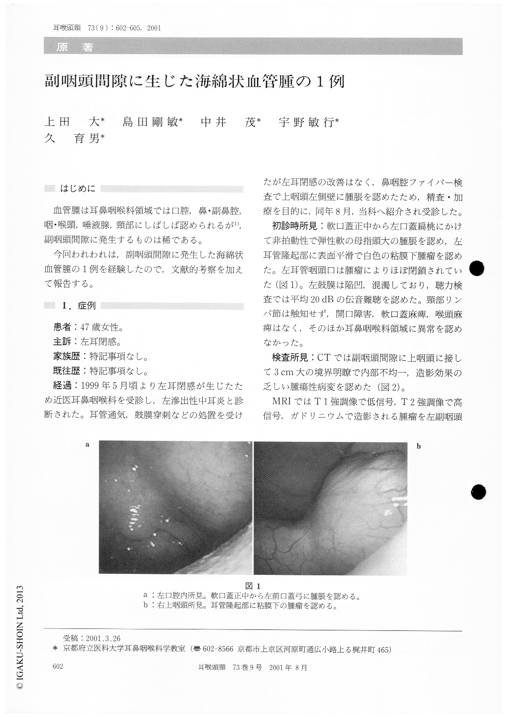
血管腫は耳鼻咽喉科領域では口腔鼻・副鼻腔,咽・喉頭,唾液腺,頸部にしばしば認められるが1),副咽頭間隙に発生するものは稀である。
今回われわれは,副咽頭間隙に発生した海綿状血管腫の1例を経験したので,文献的考察を加えて報告する。
A 47-year-old woman with a complain of 3 months'history of fullness of the left ear. The tumor was occupied in the prestyloid compartment of theleft parapharyngeal space.
Enhanced CT scan showed a clear margin and heterogenic density of the tumor. MRI of the tumor showed low intensity on T 1 weighted image and high intensity on T 2 weighted image. The cyto-logical diagnosis was blood cells.
The surgical removal of the tumor was performed by the oral approach. The pathological diagnosis was cavernous hemangioma. She showed no event-ful episode after 17 months postoperatively.
Copyright © 2001, Igaku-Shoin Ltd. All rights reserved.