Japanese
English
- 有料閲覧
PPV購入案内
1,320円 (1,200円+税10%) こちらは、94ページから98ページの文献です。購入には医書.jp本会員登録が必要です。
会員登録完了後に改めて上記「購入サイトへ」を選択してください。
または「購入サイトへ」選択後に「購入ログイン」、「新規会員登録」の順に進んでください。
会員登録について詳しくはをご確認ください。
- Abstract 文献概要
- 1ページ目 Look Inside
はじめに
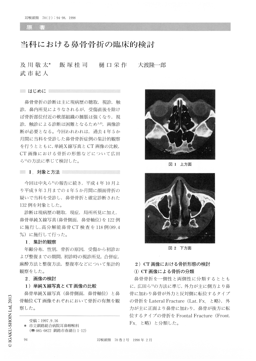
鼻骨骨折の診断は主に現病歴の聴取,視診,触診,鼻内所見によりなされるが,受傷直後を除けば骨折部位付近の軟部組織の腫脹は強くなり,視診,触診による診断は困難となるため1,2),画像診断が必要となる。今回われわれは,過去4年5か月間に当科を受診した鼻骨骨折症例の集計的観察を行うとともに,単純X線写真とCT画像の比較,CT画像における骨折の形態などについて広田ら1)の方法に準じて検討した。
One hundred thirty two patients with nasal frac-ture were diagnosed in our department from Octo-ber 1992 to March 1997. Nasal fracture was com-monly seen in teen aged males. The most frequentcauses of nasal fracture were sports.
We evaluated the computerized tomography (CT) images on three planes including upper, mid-dle and lower portions of the nasal bone. In the middle and lower portions, nasal fractures were more frequent than in upper portion.
Bilateral nasal fractures were more frequent than unilateral, and lateral fractures were more frequent than frontal.
Copyright © 1998, Igaku-Shoin Ltd. All rights reserved.