Japanese
English
- 有料閲覧
PPV購入案内
1,320円 (1,200円+税10%) こちらは、29ページから33ページの文献です。購入には医書.jp本会員登録が必要です。
会員登録完了後に改めて上記「購入サイトへ」を選択してください。
または「購入サイトへ」選択後に「購入ログイン」、「新規会員登録」の順に進んでください。
会員登録について詳しくはをご確認ください。
- Abstract 文献概要
- 1ページ目 Look Inside
はじめに
鼻科領域の手術はマイクロサージェリーが普及した現在においても裸眼手術が主流である1)。ファイバースコープはblindnessの解消,深達性で優れ,手術にも積極的に導入されつつある2〜4)。一方,顕微鏡手術は便宜さの点でファイバースコープに劣るが,安全性,術野の確保,拡大率,全身麻酔の適応などの点で優れている5〜7)。これらの長所は視神経管走行をめぐる解剖学的バリエーション8〜10)や重篤合併症1,11)を考慮すると,後部副鼻腔手術に適している。
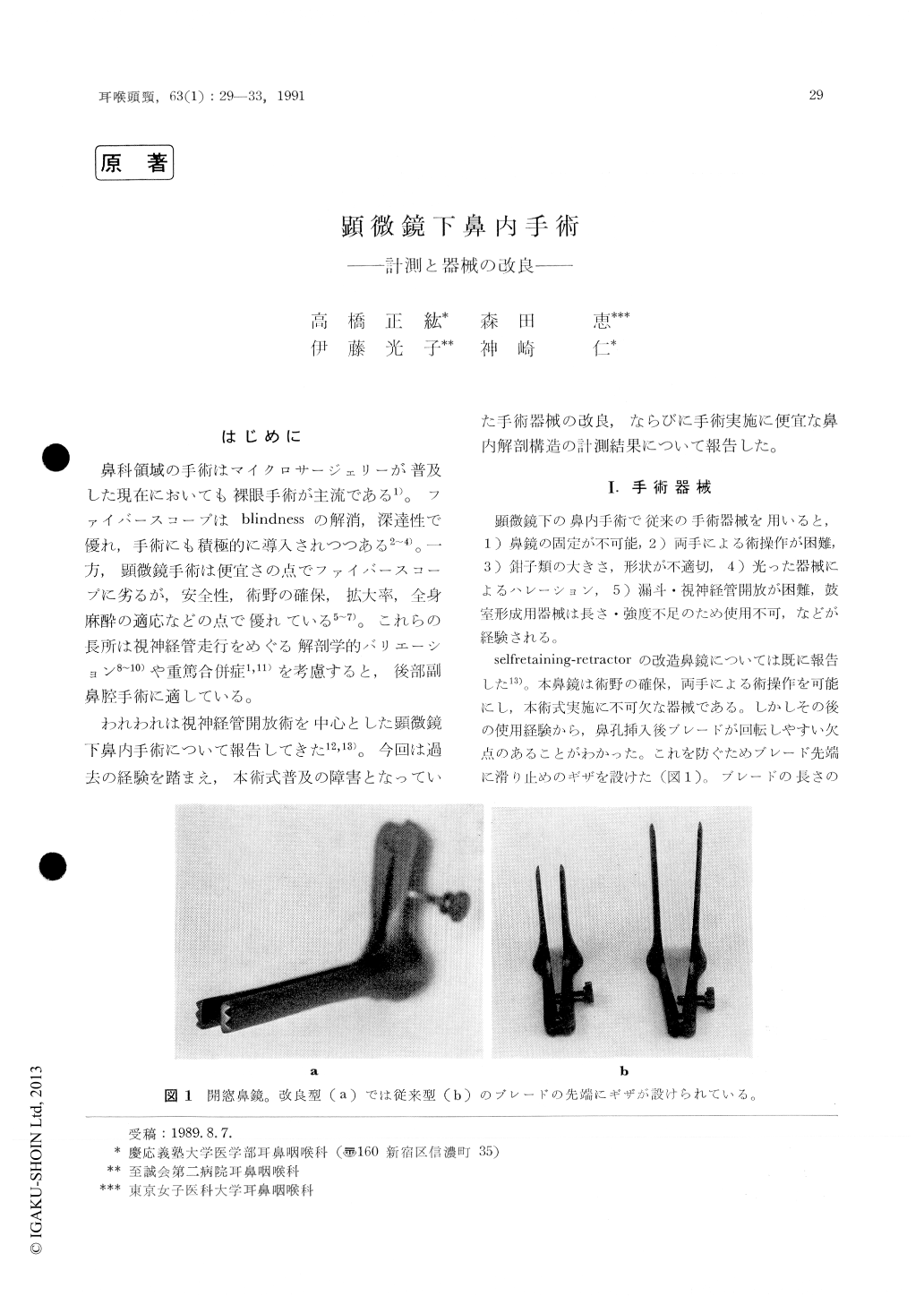
われわれは視神経管開放術を中心とした顕微鏡下鼻内手術について報告してきた12,13)。今回は過去の経験を踏まえ,本術式普及の障害となっていた手術器械の改良,ならびに手術実施に便宜な鼻内解剖構造の計測結果について報告した。
We reported surgical instruments for microscopic intranasal operation such as a self-retaining ret-ractor speculum, small forceps and stripping knives for optic nerve decompression. Furthermore, we measured the lengths from the nostril to traget structures and the angle between a view axis and the nose back on lateral view of x-ray films in 95 persons which were taken with a piece of solder put on patient's nose back. The depth to each of the target structures (the roof of the ethmoid cavity, optic nerve canal, the frontal and rear walls of the sphenoid cavity, the nasopharyngeal wall) increase with aging until a mid-teen age. In contrast, the angle of view axis decrease with aging. We showed predicted values of the depth and angle at the age of 3, 7, 10 and 16 years.
Copyright © 1991, Igaku-Shoin Ltd. All rights reserved.