Japanese
English
- 有料閲覧
- Abstract 文献概要
- 1ページ目 Look Inside
はじめに
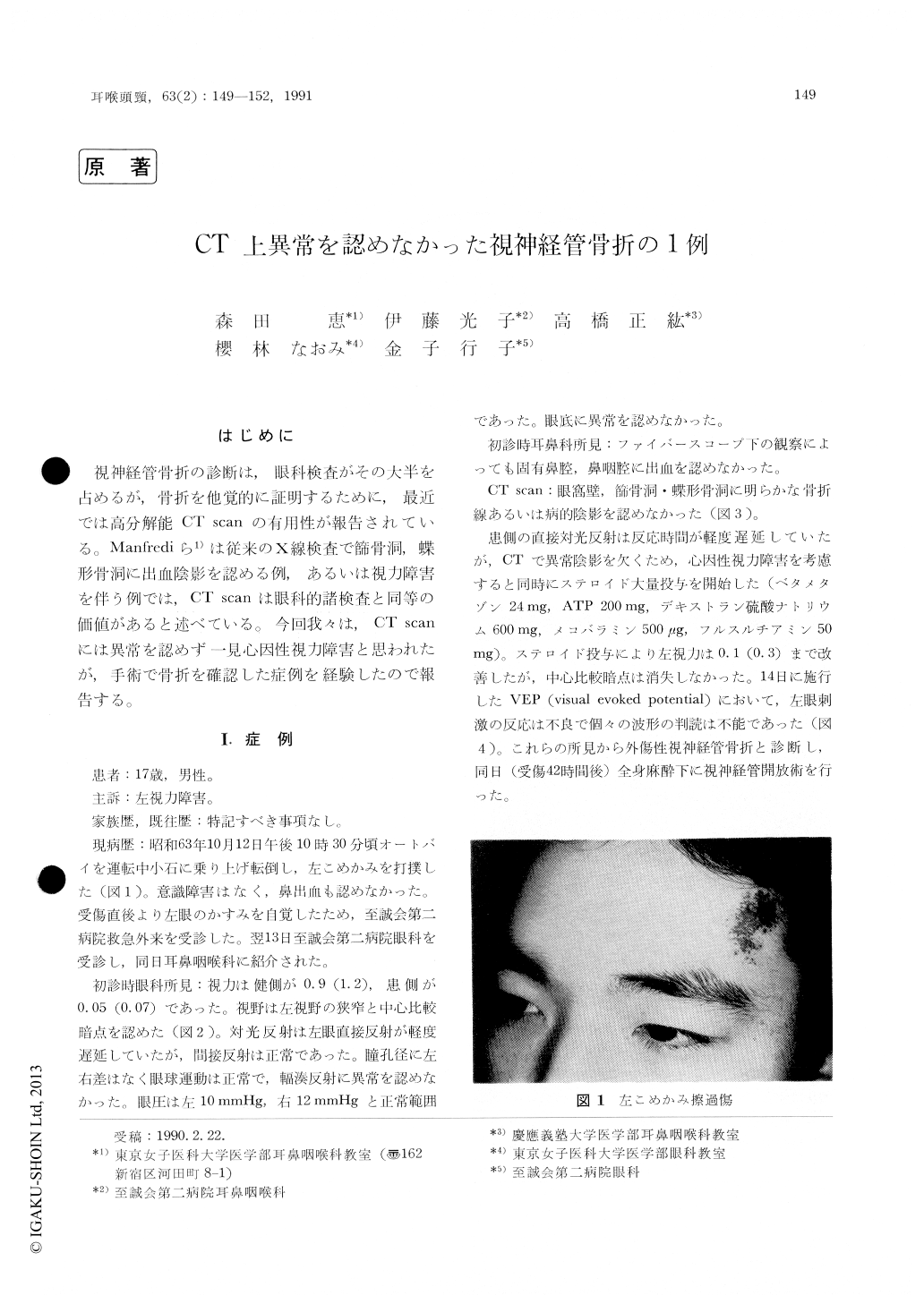
視神経管骨折の診断は,眼科検査がその大半を占めるが,骨折を他覚的に証明するために,最近では高分解能CT scanの有用性が報告されている。Manfrediら1)は従来のX線検査で篩骨洞,蝶形骨洞に出血陰影を認める例,あるいは視力障害を伴う例では,CT scanは眼科的諸検査と同等の価値があると述べている。今回我々は,CT scanには異常を認めず一見心因性視力障害と思われたが,手術で骨折を確認した症例を経験したので報告する。
A 17-year-old boy noticed visual disturbance on the left eye immediately after forehead trauma. He presented neither epistaxis, nor optic canal fracture, and pathological shadow in sinuses on x-ray films. He demonstrated a sign of Marcus-Gunn pupil on the left eye. VEP showed abnormal pattern. Surgery showed optic canal fracture. After surgery, visual acuity recovered from 0.05 to 0.5. Direct light reflex and VEP were useful to diag-nose small optic canal fracture which was hard to be detected by CT scan.

Copyright © 1991, Igaku-Shoin Ltd. All rights reserved.


