Japanese
English
- 有料閲覧
- Abstract 文献概要
- 1ページ目 Look Inside
I.はじめに
外傷性視神経障害はその病態に対する解釈はさまざまである1〜3)が,原則的に外科的治療のよい適応である。視神経管開放術にはアプローチの異なる種々の方法があるが,一般には鼻外法2)および経上顎洞法4,5)が多く行われている。鼻内法6)は視角視野の点で最も優れたアプローチといわれている4,7)が,技術的理由から必ずしも多く行われていない。
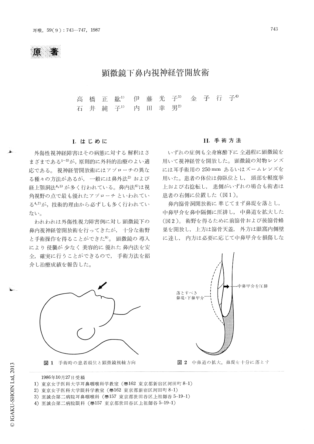
われわれは外傷性視力障害例に対し顕微鏡下の鼻内視神経管開放術を行ってきたが,十分な術野と手術操作を得ることができた8)。顕微鏡の導入により侵襲が少なく美容的に優れた鼻内法を安全,確実に行うことができるので,手術方法を紹介し治療成績を報告した。
Microscopic endonasal decompression of the optic nerve was performed in patients with optic nerve injuries. After widely opening the ethmoid and sphenoid cavity along the orbital funnel, we could detect the optic bony canal wall between the end of the orbital funnel and the contralateral sphenoid cavity. The keys to successful operation were to appropriately adjust the angle between a microscope and patient's head, to early detect the anterior sphenoid wall and to open the funnel previous to the optic canal.
All five patients treated with this method including a 7-year-old girl showed an immediate to early recovery after operation. Endonasal approach under a microscope proved to be safe and reliable as well as cosmetically favorable.

Copyright © 1987, Igaku-Shoin Ltd. All rights reserved.


