Japanese
English
- 有料閲覧
PPV購入案内
1,320円 (1,200円+税10%) こちらは、1071ページから1074ページの文献です。購入には医書.jp本会員登録が必要です。
会員登録完了後に改めて上記「購入サイトへ」を選択してください。
または「購入サイトへ」選択後に「購入ログイン」、「新規会員登録」の順に進んでください。
会員登録について詳しくはをご確認ください。
- Abstract 文献概要
- 1ページ目 Look Inside
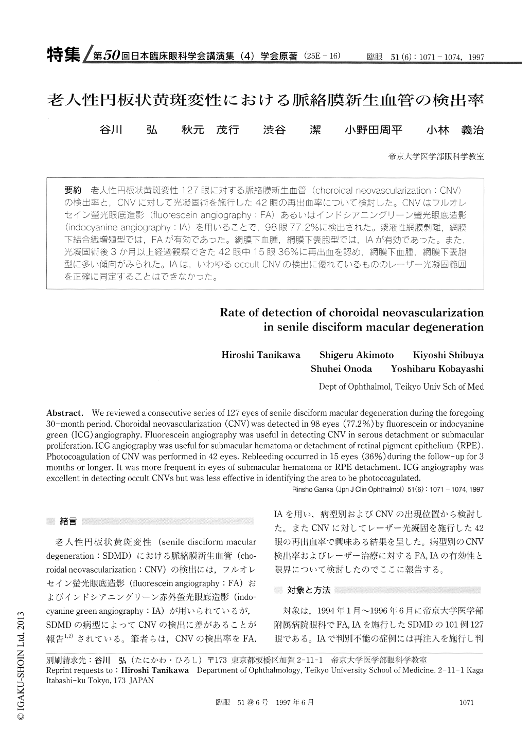
(25E-16) 老人性円板状黄斑変性127眼に対する脈絡膜新生血管(choroidal neovascularization:CNV)の検出率と,CNVに対して光凝固術を施行した42眼の再出血率について検討した。CNVはフルオレセイン螢光眼底造影(fluorescein angiography:FA)あるいはインドシアニングリーン螢光眼底造影(indocyanine angiography:IA)を用いることで198眼77.2%に検出された。漿液性網膜剥離,網膜下結合織増殖型では,FAが有効であった。網膜下血腫,網膜下嚢胞型では,IAが有効であった。また,光凝固術後3か月以上経過観察できた42眼中15眼36%に再出血を認め,網膜下血腫,網膜下嚢胞型に多い傾向がみられた。IAは,いわゆるoccult CNVの検出に優れているもののレーザー光凝固範囲を正確に同定することはできなかった。
We reviewed a consecutive series of 127 eyes of senile disciform macular degeneration during the foregoing 30-month period. Choroidal neovascularization (CNV) was detected in 98 eyes (77.2%) by fluorescein or indocyanine green (ICG) angiography. Fluorescein angiography was useful in detecting CNV in serous detachment or submacular proliferation. ICG angiography was useful for submacular hematoma or detachment of retinal pigment epithelium (RPE). Photocoagulation of CNV was performed in 42 eyes. Rebleeding occurred in 15 eyes (36%) during the follow-up for 3 months or longer. It was more frequent in eyes of submacular hematoma or RPE detachment. ICG angiography was excellent in detecting occult CNVs but was less effective in identifying the area to be photocoagulated.
Copyright © 1997, Igaku-Shoin Ltd. All rights reserved.