Japanese
English
- 有料閲覧
- Abstract 文献概要
- 1ページ目 Look Inside
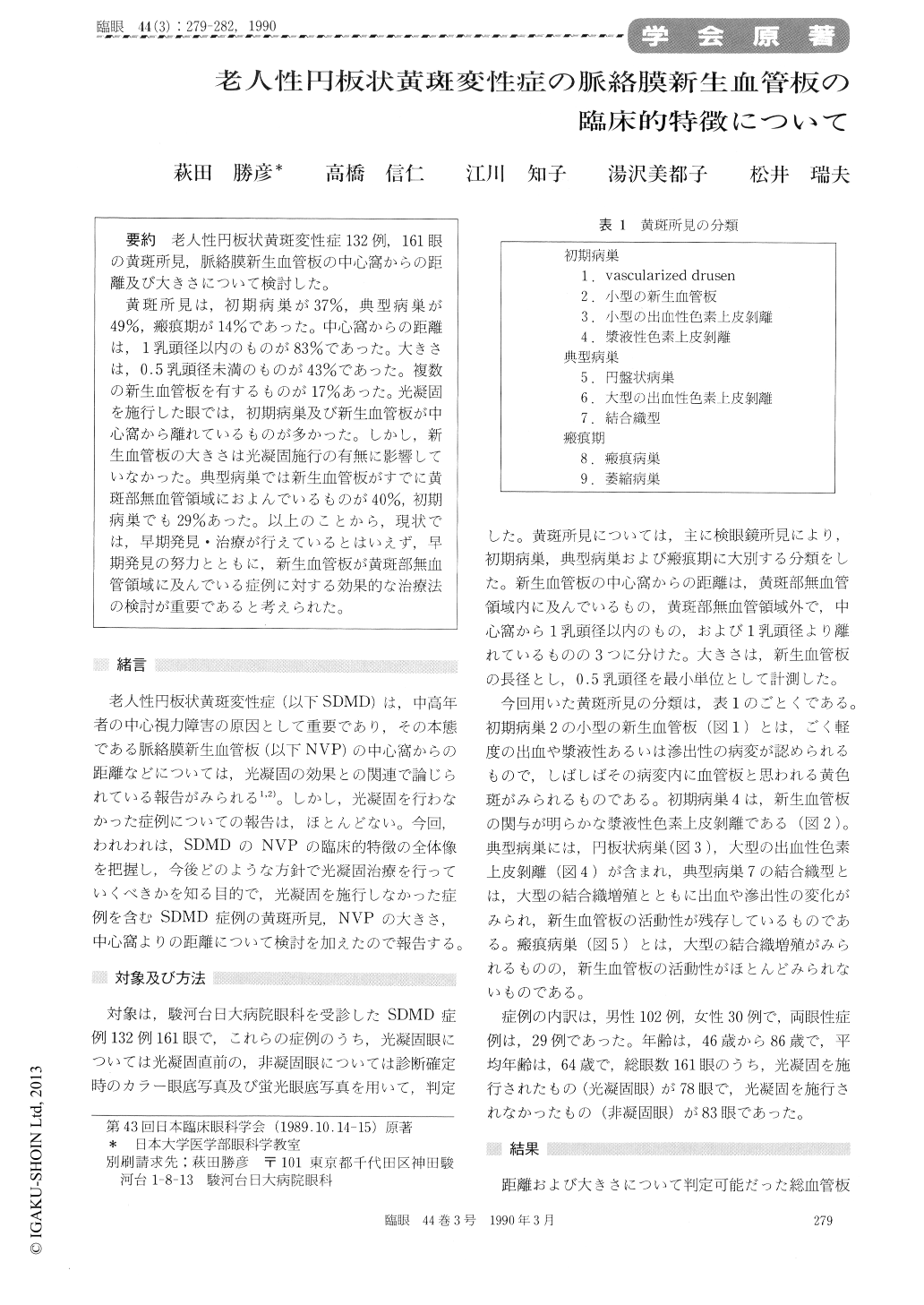
老人性円板状黄斑変性症132例,161眼の黄斑所見,脈絡膜新生血管板の中心窩からの距離及び大きさについて検討した。
黄斑所見は,初期病巣が37%,典型病巣が49%,瘢痕期が14%であった。中心窩からの距離は,1乳頭径以内のものが83%であった。大きさは,0.5乳頭径未満のものが43%であった。複数の新生血管板を有するものが17%あった。光凝固を施行した眼では,初期病巣及び新生血管板が中心窩から離れているものが多かった。しかし,新生血管板の大きさは光凝固施行の有無に影響していなかった。典型病巣では新生血管板がすでに黄斑部無血管領域におよんでいるものが40%,初期病巣でも29%あった。以上のことから,現状では,早期発見・治療が行えているとはいえず,早期発見の努力とともに,新生血管板が黄斑部無血管領域に及んでいる症例に対する効果的な治療法の検討が重要であると考えられた。
We reviewed a series of 161 eyes with senile disciform macular degeneration (SDMD) regarding the angiographic findings of subretinal neovas-cularization (SRN) and ophthalmoscopic findings. We paid particular attention to the size, location and the stage of SRNs as related to pontential photocoagulation.
Out of the whole series, 60 eyes (37%) were assigned to the early stage, 79 (49%) to typical active stage and 22 (14%) to the cicatricial stage.Out of a total of 166 SRNs, 137 (83%) were located within one disc diameter from center of the fovea. The SRNs were smaller than 0.5 disc diameter in 71 eyes (43%). In eyes with typical active stage, 35 of 88 SRNs (40%) were located within the foveal avascular zone (FAZ). In eyes with early stage of the lesion, 21 of 73 SRNs (29%) were located within the FAZ.
The finding seemed to imply that an utmost effort is to be paid in early detection of SDMD and to develop a therapeutic modality to treat SRNs located within the FAZ.

Copyright © 1990, Igaku-Shoin Ltd. All rights reserved.


