Japanese
English
- 有料閲覧
PPV購入案内
1,320円 (1,200円+税10%) こちらは、381ページから384ページの文献です。購入には医書.jp本会員登録が必要です。
会員登録完了後に改めて上記「購入サイトへ」を選択してください。
または「購入サイトへ」選択後に「購入ログイン」、「新規会員登録」の順に進んでください。
会員登録について詳しくはをご確認ください。
- Abstract 文献概要
- 1ページ目 Look Inside
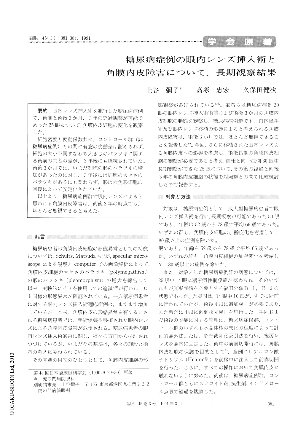
眼内レンズ挿入術を施行した糖尿病症例で,術前と術後3か月,3年の経過観察が可能であった25眼について,角膜内皮細胞の変化を観察した。
細胞密度と変動係数共に,コントロール群(非糖尿病症例)との間に有意の変動差は認められず,細胞の大小不同すなわち大きさのバラツキに関する術前の両者の差が,3年後にも継続されていた。術後3か月では,いまだ細胞の形のバラツキの増加があったのに対し,3年後には細胞の大きさのバラツキがあるにも関わらず,形は六角形細胞の回復によって安定化されていた。
以上より,糖尿病症例群で眼内レンズによると思われる角膜内皮障害は,術後3年の時点でも,ほとんど無視できると考えた。
We evaluated the corneal endothelium in 25 diabetic eyes 3 years after extracapsular cataract extraction or phacoemulsification with posterior chamber lens implantation. The corneal endoth-elium was documented with contact lens of Eisner and processed by graph pen digitizer. Another series of 50 nondiabetic eyes served as control. No statistical difference in the cell density was foundbetween the diabetic subjects and the nondiabetic subjects. The diabetic subjects had a significantly higher (coefficient of variation (0.34±0.06) than the controls (0.31±0.05). The difference from presurgical state was essentially similar between both. There was a significant recovery in the per-centage of hexagonal cells in them. These results indicate the endothelial cells are minimally affected by posterior chamber lens implantation in diabetic eyes.
Copyright © 1991, Igaku-Shoin Ltd. All rights reserved.