Japanese
English
- 有料閲覧
- Abstract 文献概要
- 1ページ目 Look Inside
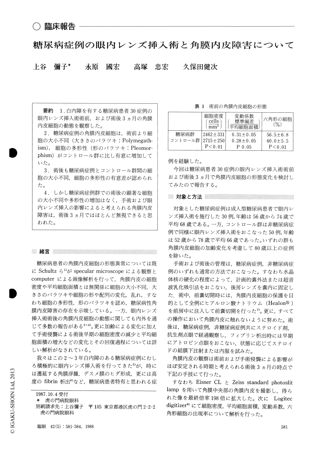
1.白内障を有する糖尿病患者30症例の眼内レンズ挿入術術前,および術後3カ月の角膜内皮細胞の動態を観察した.
2.糖尿病症例の角膜内皮細胞は,術前より細胞の大小不同(大きさのバラツキ:Polymegath-ism),細胞の多形性(形のバラツキ:Pleomor-phism)がコントロール群に比し有意に増加していた.
3.術後も糖尿病症例とコントロール群間の細胞の大小不同,細胞の多形性の有意差が認められた.
4.しかし糖尿病症例群での術後の顕著な細胞の大小不同や多形性の増加はなく,手術および眼内レンズ挿入の影響によると考えられる角膜内皮障害は,術後3カ月ではほとんど無視できると思われた.
We evaluated the corneal endothelium in 30 diabetic eyes before and 3 months after extracap-sular cataract extraction or phacoemulsification with posterior intraocular lens implantation. The corneal endothelium was documented with contact lens of Eisner and processed by graph pen digitizer. Another series of 50 non-diabetic eyes served as control.
Prior to surgery, the corneal endothelium in diabetic eyes showed significantly decreased cell density, increased coefficient of variation orpolymegathism, and decrease in hexagonal cells or pleomorphism when compared with controls.
After surgery, the difference from presurgical state was essentially similar in diabetic and non -diabetic groups.
In particular, the polymegathism and pleomorphism in the diabetic eyes failed to show marked increase following surgery. The findings show that corneal endothelium in diabetic eyes is minimally affected by extracap-sular cataract extraction and posterior intraocular lens implantation under careful surgical manage-ment.
Rinsho Ganka (Jpn J Clin Ophthalmol) 42(5) : 581-584, 1988

Copyright © 1988, Igaku-Shoin Ltd. All rights reserved.


