Japanese
English
- 有料閲覧
PPV購入案内
1,320円 (1,200円+税10%) こちらは、1483ページから1487ページの文献です。購入には医書.jp本会員登録が必要です。
会員登録完了後に改めて上記「購入サイトへ」を選択してください。
または「購入サイトへ」選択後に「購入ログイン」、「新規会員登録」の順に進んでください。
会員登録について詳しくはをご確認ください。
- Abstract 文献概要
- 1ページ目 Look Inside
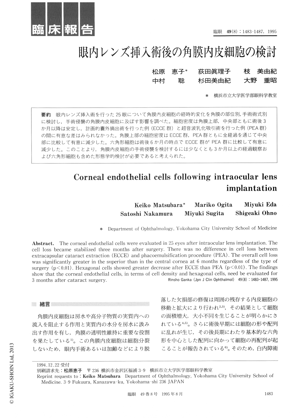
眼内レンズ挿入術を行った25眼について角膜内皮細胞の経時的変化を角膜の部位別,手術術式別に検討し,手術侵襲の角膜内皮細胞に及ぼす影響を調べた。細胞密度は角膜上部,中央部ともに術後3か月以降は安定し,計画的嚢外摘出術を行った例(ECCE群)と超音波乳化吸引術を行った例(PEA群)の間に有意な差はみられなかった。角膜上部の細胞密度はECCE群,PEA群ともに全経過を通じて中央部に比較して有意に減少した。六角形細胞は術後6か月の時点でECCE群がPEA群に比較して有意に減少した。このことより,角膜内皮細胞の手術侵襲を検討するには少なくとも3か月以上の経過観察および六角形細胞も含めた形態学的検討が必要であると考えられた。
The corneal endothelial cells were evaluated in 25 eyes after intraocular lens implantation. Thecell loss became stabilized three months after surgery. There was no difference in cell loss betweenextracapsular cataract extraction (ECCE) and phacoemulsification procedure (PEA). The overall cell losswas significantly greater in the superior than in the central cornea at 6 months regardless of the type ofsurgery (p<0.01). Hexagonal cells showed greater decrease after ECCE than PEA (p<0.01). The findingsshow that the corneal endothelial cells, in terms of cell density and hexagonal cells, need be evaluated for3 months after cataract surgery.
Copyright © 1995, Igaku-Shoin Ltd. All rights reserved.