Japanese
English
- 有料閲覧
PPV購入案内
1,320円 (1,200円+税10%) こちらは、421ページから424ページの文献です。購入には医書.jp本会員登録が必要です。
会員登録完了後に改めて上記「購入サイトへ」を選択してください。
または「購入サイトへ」選択後に「購入ログイン」、「新規会員登録」の順に進んでください。
会員登録について詳しくはをご確認ください。
- Abstract 文献概要
- 1ページ目 Look Inside
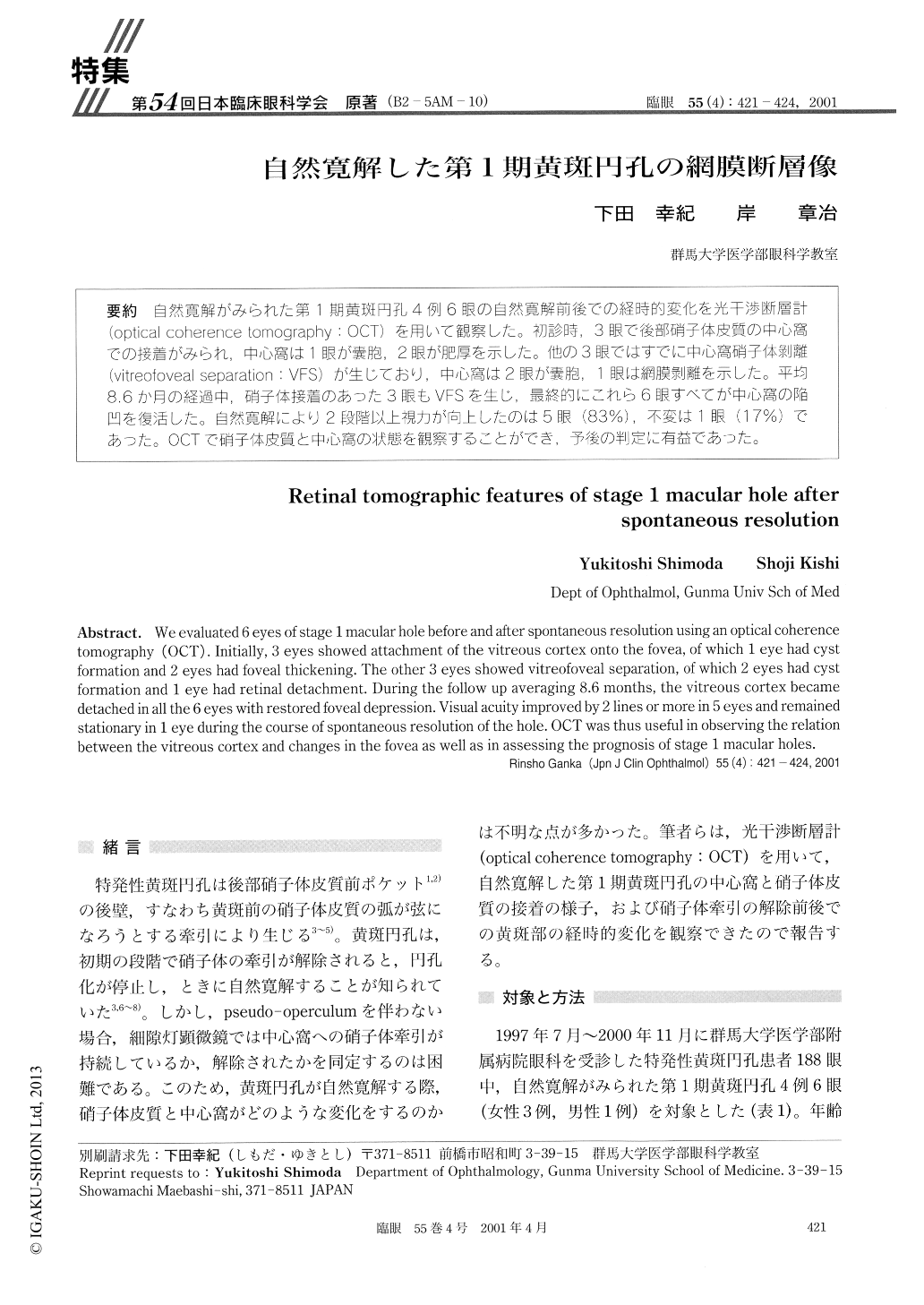
自然寛解がみられた第1期黄斑円孔4例6眼の自然寛解前後での経時的変化を光干渉断層計(optical coherence tomography:OCT)を用いて観察した。初診時,3眼で後部硝子体皮質の中心窩での接着がみられ,中心窩は1眼が嚢胞,2眼が肥厚を示した。他の3眼ではすでに中心窩硝子体剥離(vitreofoveal separation:VFS)が生じており,中心窩は2眼が嚢胞,1眼は網膜剥離を示した。平均8.6か月の経過中,硝子体接着のあった3眼もVFSを生じ,最終的にこれら6眼すべてが中心窩の陥凹を復活した。自然寛解により2段階以上視力が向上したのは5眼(83%),不変は1眼(17%)であった。OCTで硝子体皮質と中心窩の状態を観察することができ,予後の判定に有益であった。
We evaluated 6 eyes of stage 1 macular hole before and after spontaneous resolution using an optical coherence tomography (OCT). Initially, 3 eyes showed attachment of the vitreous cortex onto the fovea, of which 1 eye had cyst formation and 2 eyes had foveal thickening. The other 3 eyes showed vitreofoveal separation, of which 2 eyes had cyst formation and 1 eye had retinal detachment. During the follow up averaging 8.6 months, the vitreous cortex became detached in all the 6 eyes with restored foveal depression. Visual acuity improved by 2 lines or more in 5 eyes and remained stationary in 1 eye during the course of spontaneous resolution of the hole. OCT was thus useful in observing the relation between the vitreous cortex and changes in the fovea as well as in assessing the prognosis of stage 1 macular holes.
Copyright © 2001, Igaku-Shoin Ltd. All rights reserved.