Japanese
English
- 有料閲覧
- Abstract 文献概要
- 1ページ目 Look Inside
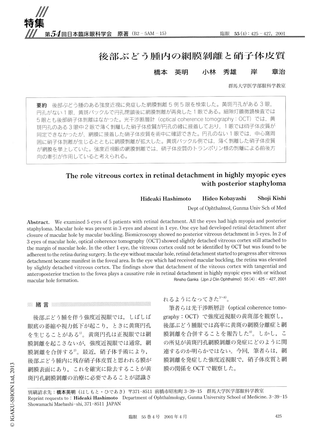
後部ぶどう腫のある強度近視に発症した網膜剥離5例5眼を検索した。黄斑円孔がある3眼,円孔がない1眼,黄斑バックルで円孔閉鎖後に網膜剥離が再発した1眼である。細隙灯顕微鏡検査では5眼とも後部硝子体剥離はなかった。光干渉断層計(optical coherence tomography:OCT)では,黄斑円孔のある3眼中2眼で薄く剥離した硝子体皮質が円孔の縁に接着しており,1眼では硝子体皮質が同定できなかったが,網膜に接着した硝子体皮質を術中に確認できた。円孔のない1眼では,中心窩周囲に硝子体剥離が生じるとともに網膜剥離が拡大した。黄斑バックル例では,薄く剥離した硝子体皮質が網膜を挙上していた。強度近視眼の網膜剥離では、硝子体皮質のトランポリン様の剥離による前後方向の牽引が作用していると考えられる。
We examined 5 eyes of 5 patients with retinal detachment. All the eyes had high myopia and posterior staphyloma. Macular hole was present in 3 eyes and absent in 1 eye. One eye had developed retinal detachment after closure of macular hole by macular buckling. Biomicroscopy showed no posterior vitreous detachment in 5 eyes. In 2 of 3 eyes of macular hole, optical coherence tomography (OCT) showed slightly detached vitreous cortex still attached to the margin of macular hole. In the other 1 eye, the vitreous cortex could not be identified by OCT but was found to be adherent to the retina during surgery. In the eye without macular hole, retinal detachment started to progress after vitreous detachment became manifest in the foveal area. In the eye which had received macular buckling, the retina was elevated by slightly detached vitreous cortex. The findings show that detachment of the viteous cortex with tangential and anteroposterior traction to the fovea plays a causative role in retinal detachment in highly myopic eyes with or without macular hole formation.

Copyright © 2001, Igaku-Shoin Ltd. All rights reserved.


