Japanese
English
- 有料閲覧
PPV購入案内
1,320円 (1,200円+税10%) こちらは、685ページから687ページの文献です。購入には医書.jp本会員登録が必要です。
会員登録完了後に改めて上記「購入サイトへ」を選択してください。
または「購入サイトへ」選択後に「購入ログイン」、「新規会員登録」の順に進んでください。
会員登録について詳しくはをご確認ください。
- Abstract 文献概要
- 1ページ目 Look Inside
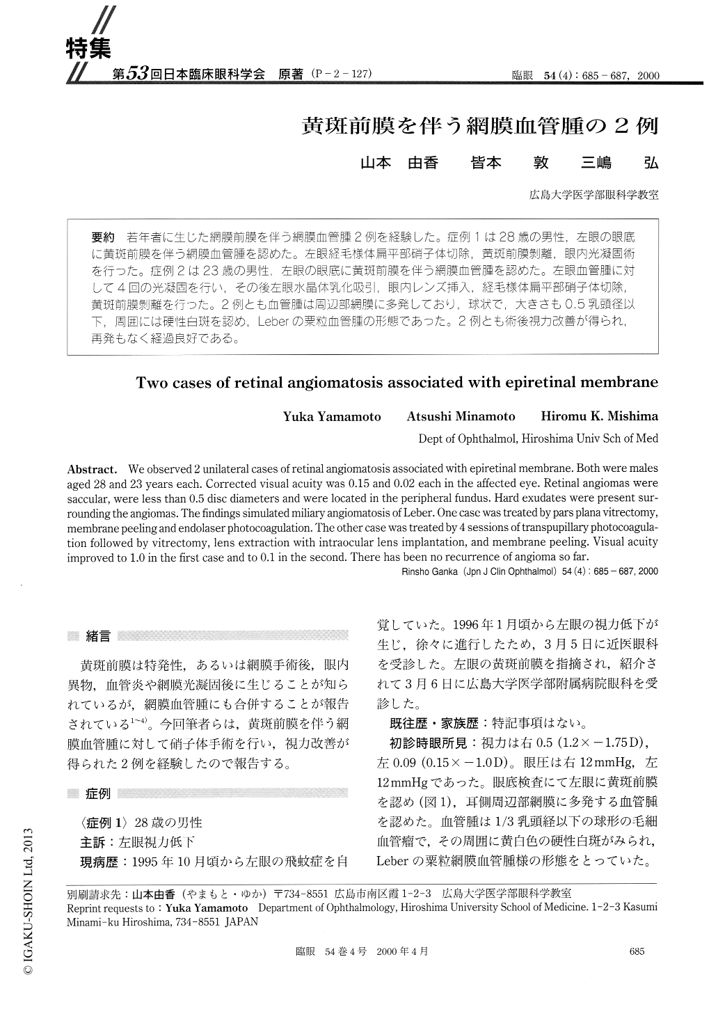
(P-2-127) 若年者に生じた網膜前膜を伴う網膜血管腫2例を経験した。症例1は28歳の男性,左眼の眼底に黄斑前膜を伴う網膜血管腫を認めた。左眼経毛様体扁平部硝子体切除,黄斑前膜剥離,眼内光凝固術を行った。症例2は23歳の男性,左眼の眼底に黄斑前膜を伴う網膜血管腫を認めた。左眼血管腫に対して4回の光凝固を行い,その後左眼水晶体乳化吸引,眼内レンズ挿入,経毛様体扁平部硝子体切除,黄斑前膜剥離を行った。2例とも血管腫は周辺部網膜に多発しており,球状で,大きさも0.5乳頭径以下,周囲には硬性白斑を認め,Leberの粟粒血管腫の形態であった。2例とも術後視力改善が得られ,再発もなく経過良好である。
We observed 2 unilateral cases of retinal angiomatosis associated with epiretinal membrane. Both were males aged 28 and 23 years each. Corrected visual acuity was 0.15 and 0.02 each in the affected eye. Retinal angiomas were saccular, were less than 0.5 disc diameters and were located in the peripheral fundus. Hard exudates were present sur-rounding the angiomas. The findings simulated miliary angiomatosis of Leber. One case was treated by pars plana vitrectomy, membrane peeling and endolaser photocoagulation. The other case was treated by 4 sessions of transpupillary photocoagula-tion followed by vitrectomy, lens extraction with intraocular lens implantation, and membrane peeling. Visual acuity improved to 1.0 in the first case and to 0.1 in the second. There has been no recurrence of angioma so far.
Copyright © 2000, Igaku-Shoin Ltd. All rights reserved.