Japanese
English
- 有料閲覧
PPV購入案内
1,320円 (1,200円+税10%) こちらは、680ページから684ページの文献です。購入には医書.jp本会員登録が必要です。
会員登録完了後に改めて上記「購入サイトへ」を選択してください。
または「購入サイトへ」選択後に「購入ログイン」、「新規会員登録」の順に進んでください。
会員登録について詳しくはをご確認ください。
- Abstract 文献概要
- 1ページ目 Look Inside
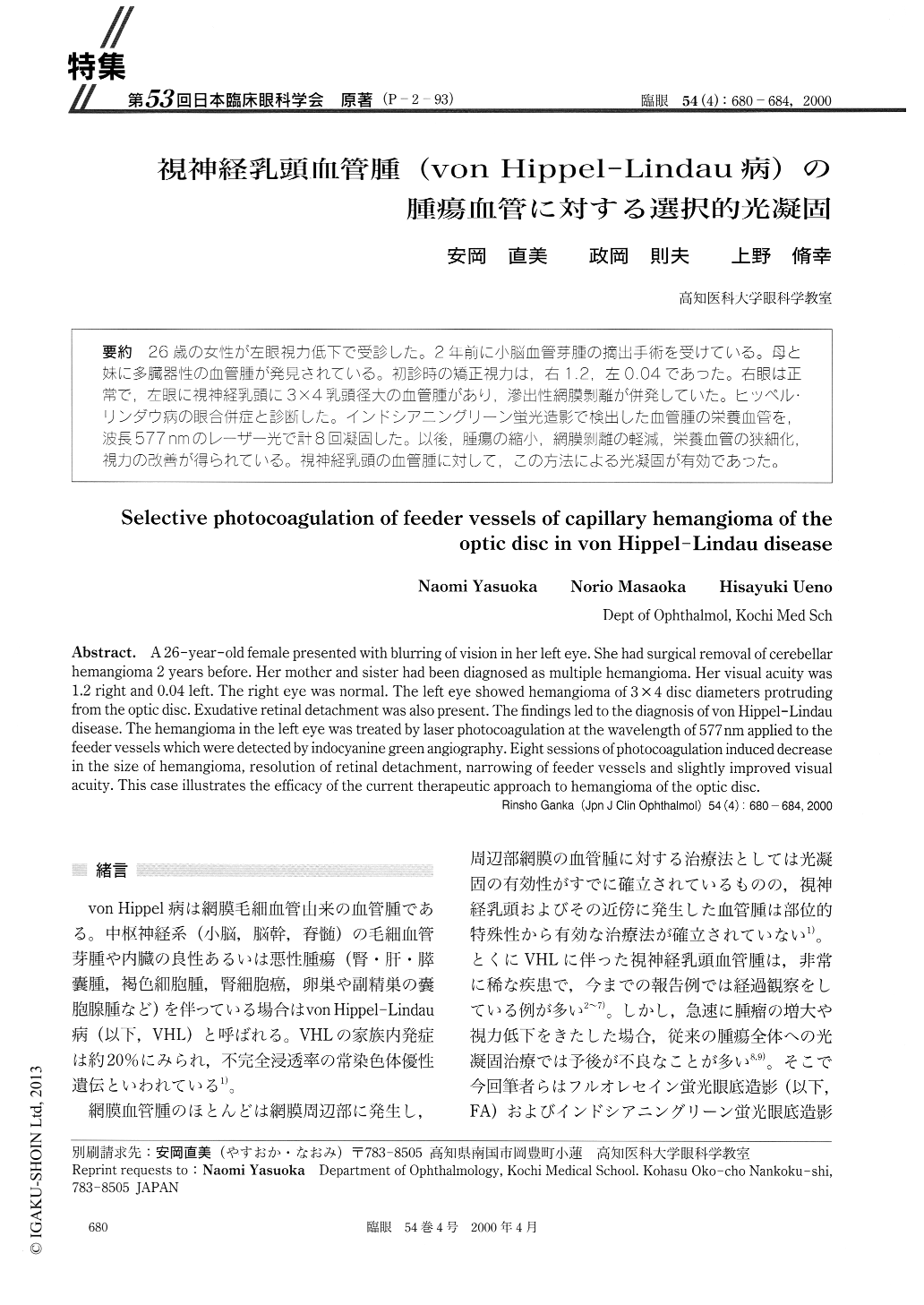
(P-2-93) 26歳の女性が左眼視力低下で受診した。2年前に小脳血管芽腫の摘出手術を受けている。母と妹に多臓器性の血管腫が発見されている。初診時の矯正視力は,右1.2,左0.04であった。右眼は正常で,左眼に視神経乳頭に3×4乳頭径大の血管腫があり,滲出性網膜剥離が併発していた。ヒッペルリンダウ病の眼合併症と診断した。インドシアニングリーン蛍光造影で検出した血管腫の栄養血管を,波長577nmのレーザー光で計8回凝固した。以後,腫瘍の縮小,網膜剥離の軽減,栄養血管の狭細化,視力の改善が得られている。視神経乳頭の血管腫に対して,この方法による光凝固が有効であった。
A 26-year-old female presented with blurring of vision in her left eye. She had surgical removal of cerebellar hemangioma 2 years before. Her mother and sister had been diagnosed as multiple hemangioma. Her visual acuity was 1.2 right and 0.04 left. The right eye was normal. The left eye showed hemangioma of 3 ×4 disc diameters protruding from the optic disc. Exudative retinal detachment was also present. The findings led to the diagnosis of von Hippel-Lindau disease. The hemangioma in the left eye was treated by laser photocoagulation at the wavelength of 577nm applied to the feeder vessels which were detected by indocyanine green angiography. Eight sessions of photocoagulation induced decrease in the size of hemangioma, resolution of retinal detachment, narrowing of feeder vessels and slightly improved visual acuity. This case illustrates the efficacy of the current therapeutic approach to hemangioma of the optic disc.
Copyright © 2000, Igaku-Shoin Ltd. All rights reserved.