Japanese
English
- 有料閲覧
PPV購入案内
1,320円 (1,200円+税10%) こちらは、967ページから970ページの文献です。購入には医書.jp本会員登録が必要です。
会員登録完了後に改めて上記「購入サイトへ」を選択してください。
または「購入サイトへ」選択後に「購入ログイン」、「新規会員登録」の順に進んでください。
会員登録について詳しくはをご確認ください。
- Abstract 文献概要
- 1ページ目 Look Inside
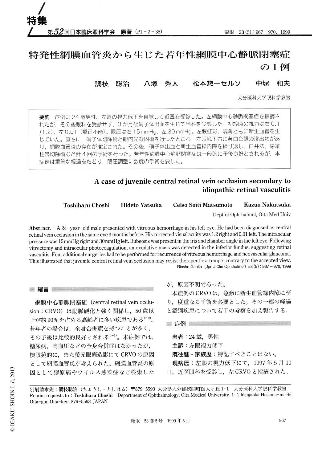
(P1-2-38) 症例は24歳男性。左眼の視力低下を自覚して近医を受診した。左網膜中心静脈閉塞症を指摘されたが,その後眼科を受診せず,3か月後硝子体出血を生じて当科を受診した。初診時の視力は右0.1(1.2),左0.01(矯正不能)。眼圧は右15mmHg,左30mmHg。左眼虹彩,隅角ともに新生血管を生じていた。直ちに,硝子体切除術と眼内光凝固術を行ったところ,左眼底下方に黄白色調の滲出物があり,網膜血管炎の存在が推定された。その後,硝子体出血と新生血管緑内障を繰り返し,臼井法,線維柱帯切除術など計4回の手術を行った。若年性網膜中心静脈閉塞症は一般的に予後良好とされるが,本症例は重篤な経過をたどり,眼圧調整に数度の手術を要した。
A 24-year-old male presented with vitreous hemorrhage in his left eye. He had been diagnosed as central retinal vein occlusion in the same eye 3 months before. His corrected visual acuity was 1.2 right and 0.01 left. The intraocular pressure was 15 mmHg right and 30 mmHg left. Rubeosis was present in the iris and chamber angle in the left eye. Following vitrectomy and intraocular photocoagulation, an exudative mass was detected in the inferior fundus, suggesting retinal vasculitis. Four additional surgeries had to be performed for recurrence of vitreous hemorrhage and neovascular glaucoma. This illustrated that juvenile central retinal vein occlusion may resist therapeutic attempts contrary to the accepted view.
Copyright © 1999, Igaku-Shoin Ltd. All rights reserved.