Japanese
English
- 有料閲覧
PPV購入案内
1,320円 (1,200円+税10%) こちらは、1234ページから1238ページの文献です。購入には医書.jp本会員登録が必要です。
会員登録完了後に改めて上記「購入サイトへ」を選択してください。
または「購入サイトへ」選択後に「購入ログイン」、「新規会員登録」の順に進んでください。
会員登録について詳しくはをご確認ください。
- Abstract 文献概要
- 1ページ目 Look Inside
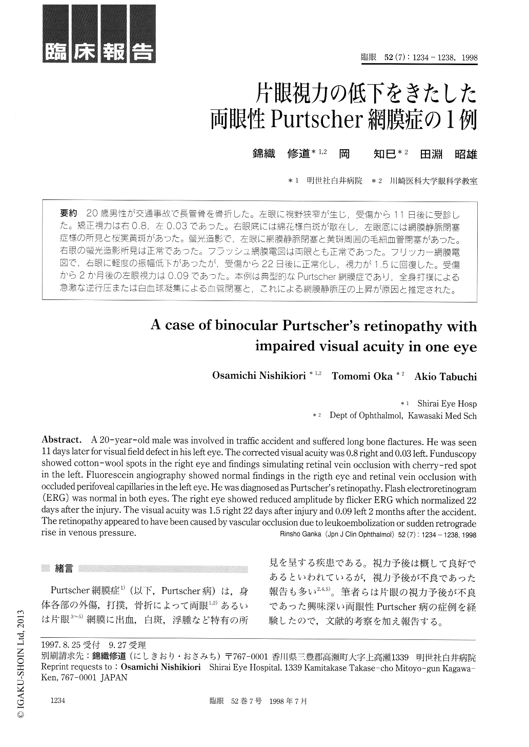
20歳男性が交通事故で長管骨を骨折した。左眼に視野狭窄が生じ,受傷から11日後に受診した。矯正視力は右0.8,左0.03であった。右眼底には綿花様白斑が散在し,左眼底には網膜静脈閉塞症様の所見と桜実黄斑があった。螢光造影で,左眼に網膜静脈閉塞と黄斑周囲の毛細血管閉塞があった。右眼の螢光造影所見は正常であった。フラッシュ網膜電図は両眼とも正常であった。フリッカー網膜電図で,右眼に軽度の振幅低下があったが,受傷から22日後に正常化し,視力が1.5に回復した。受傷から2か月後の左眼視力は0.09であった。本例は典型的なPurtscher網膜症であり,全身打撲による急激な逆行圧または白血球凝集による血管閉塞と,これによる網膜静脈圧の上昇が原因と推定された。
A 20-year-old male was involved in traffic accident and suffered long bone flactures. He was seen 11 days later for visual field defect in his left eye. The corrected visual acuity was 0.8 right and 0.03 left. Funduscopy showed cotton-wool spots in the right eye and findings simulating retinal vein occlusion with cherry-red spot in the left. Fluorescein angiography showed normal findings in the rigth eye and retinal vein occlusion with occluded perifoveal capillaries in the left eye. He was diagnosed as Purtscher's retinopathy. Flash electroretinogram (ERG) was normal in both eyes. The right eye showed reduced amplitude by flicker ERG which normalized 22 days after the injury. The visual acuity was 1.5 right 22 days after injury and 0.09 left 2 months after the accident. The retinopathy appeared to have been caused by vascular occlusion due to leukoembolization or sudden retrograde rise in venous pressure.
Copyright © 1998, Igaku-Shoin Ltd. All rights reserved.