Japanese
English
- 有料閲覧
PPV購入案内
1,320円 (1,200円+税10%) こちらは、1319ページから1323ページの文献です。購入には医書.jp本会員登録が必要です。
会員登録完了後に改めて上記「購入サイトへ」を選択してください。
または「購入サイトへ」選択後に「購入ログイン」、「新規会員登録」の順に進んでください。
会員登録について詳しくはをご確認ください。
- Abstract 文献概要
- 1ページ目 Look Inside
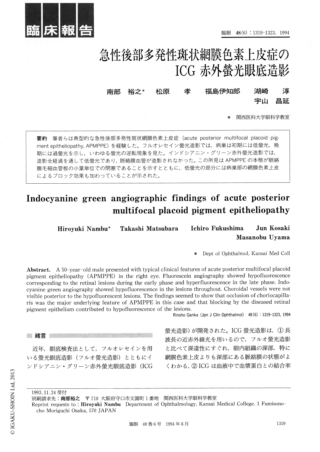
筆者らは典型的な急性後部多発性斑状網膜色素上皮症(acute posterior multifocal placoid pig—ment epitheliopathy, APMPPE)を経験した。フルオレセイン螢光造影では,病巣は初期には低螢光,晩期には過螢光を示し,いわゆる螢光の逆転現象を見た。インドシアニン・グリーン赤外螢光造影では,造影全経過を通して低螢光であり,脈絡膜血管が造影されなかった。この所見はAPMPPEの本態が脈絡膜毛細血管板の小葉単位での閉塞であることを示すとともに,低螢光の部分には病巣部の網膜色素上皮によるブロック効果も加わっていることが示された。
A 50-year-o1d male presented with typical clinical features of acute posterior multifocal placoid pigment epitheliopathy (APMPPE) in the right eye. Fluorescein angiography showed hypofluorescence corresponding to the retinal lesions during the early phase and hyperfluorescence in the late phase. Indo-cyanine green angiography showed hypofluorescence in the lesions throughout. Choroidal vessels were not visible posterior to the hypofluorescent lesions. The findings seemed to show that occlusion of choriocapilla-ris was the major underlying feature of APMPPE in this case and that blocking by the diseased retinal pigment epithelium contributed to hypofluorescence of the lesions.
Copyright © 1994, Igaku-Shoin Ltd. All rights reserved.