Japanese
English
- 有料閲覧
PPV購入案内
1,320円 (1,200円+税10%) こちらは、1513ページから1516ページの文献です。購入には医書.jp本会員登録が必要です。
会員登録完了後に改めて上記「購入サイトへ」を選択してください。
または「購入サイトへ」選択後に「購入ログイン」、「新規会員登録」の順に進んでください。
会員登録について詳しくはをご確認ください。
- Abstract 文献概要
- 1ページ目 Look Inside
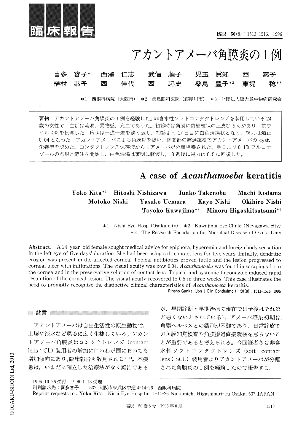
アカントアメーバ角膜炎の1例を経験した。非含水性ソフトコンタクトレンズを装用している24歳の女性で,主訴は流涙,異物感,充血であった。初診時は角膜に偽樹枝状の上皮びらんがあり,抗ウイルス剤を投与した。病状は一進一退を繰り返し,初診より17日目に白色潰瘍状となり,視力は矯正0.04となった。アカントアメーバによる角膜炎を疑い,病変部の擦過鏡検でアカントアメーバのcyst,栄養型を認めた。コンタクトレンズ保存液からもアメーバが分離培養された。翌日より0.1%フルコナゾールの点眼と静注を開始し,白色混濁は著明に軽減し,3週後に視力は0.5に回復した。
A 24-year-old female sought medical advice for epiphora, hyperemia and foreign body sensationin the left eye of five days' duration. She had been using soft contact lens for five years. Initially, dendriticerosion was present in the affected cornea. Topical antibiotics proved futile and the lesion progressed tocorneal ulcer with infiltrations. The visual acuity was now 0.04. Acanthamoeba was found in scrapings fromthe cornea and in the preservative solution of contact lens. Topical and systemic fluconazole induced rapidresolution of the corneal lesion. The visual acuity recovered to 0.5 in three weeks. This case illustrates theneed to promptly recognize the distinctive clinical characteristics of Acanthamoeba keratitis.
Copyright © 1996, Igaku-Shoin Ltd. All rights reserved.