Japanese
English
- 有料閲覧
PPV購入案内
1,320円 (1,200円+税10%) こちらは、1217ページから1220ページの文献です。購入には医書.jp本会員登録が必要です。
会員登録完了後に改めて上記「購入サイトへ」を選択してください。
または「購入サイトへ」選択後に「購入ログイン」、「新規会員登録」の順に進んでください。
会員登録について詳しくはをご確認ください。
- Abstract 文献概要
- 1ページ目 Look Inside
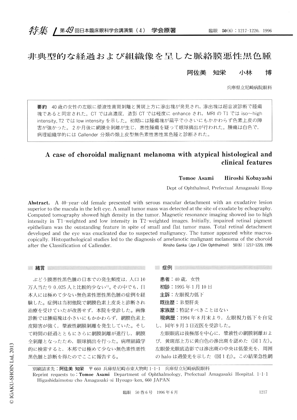
40歳の女性の左眼に漿液性黄斑剥離と黄斑上方に滲出塊が発見され,滲出塊は超音波診断で腫瘍塊であると同定された。CTでは高濃度,造影CTでは軽度にenhanceされ,MRIのT1ではiso〜high intensity,T2ではlow intensityを示した。初期には腫瘍塊が扁平で小さいにもかかわらず色素上皮の障害が強かった。2か月後に網膜全剥離が生じ,悪性腫瘍を疑って眼球摘出が行われた。腫瘍は白色で,病理組織学的にはCallender分類の類上皮型無色素性悪性黒色腫と診断された。
A 40-year-old female presented with serous macular detachment with an exudative lesion superior to the macula in the left eye. A small tumor mass was detected at the site of exudate by echography. Computed tomography showed high density in the tumor. Magnetic resonance imaging showed iso to high intensity in T1-weighted and low intensity in T2-weighted images. Initially, impaired retinal pigment epithelium was the outstanding feature in spite of small and flat tumor mass. Total retinal detachment developed and the eye was enucleated due to suspected malignancy. The tumor appeared white macros-copically. Histopathological studies led to the diagnosis of amelanotic malignant melanoma of the choroid after the Classification of Callender.
Copyright © 1996, Igaku-Shoin Ltd. All rights reserved.