Japanese
English
特集 第49回日本臨床眼科学会講演集(4)
学会原著
眼内異物の金属分析
Metallurgical analysis of intraocular foreign bodies
松野 かおり
1
,
甘利 富士夫
1
,
太田 浩一
1
,
吉村 長久
1
,
亀谷 清和
2
,
鈴木 佳代
2
Kaori Matsuno
1
,
Fujio Amari
1
,
Kouichi Ohta
1
,
Nagahisa Yoshimura
1
,
Kiyokazu Kametani
2
,
Kayo Suzuki
2
1信州大学医学部眼科学教室
2信州大学医学部総合研究室
1Dept of Ophthalmol, Shinsyu Univ Med Sch
2General Labo, Shinsyu Univ Med Sch
pp.1221-1223
発行日 1996年6月15日
Published Date 1996/6/15
DOI https://doi.org/10.11477/mf.1410904963
- 有料閲覧
- Abstract 文献概要
- 1ページ目 Look Inside
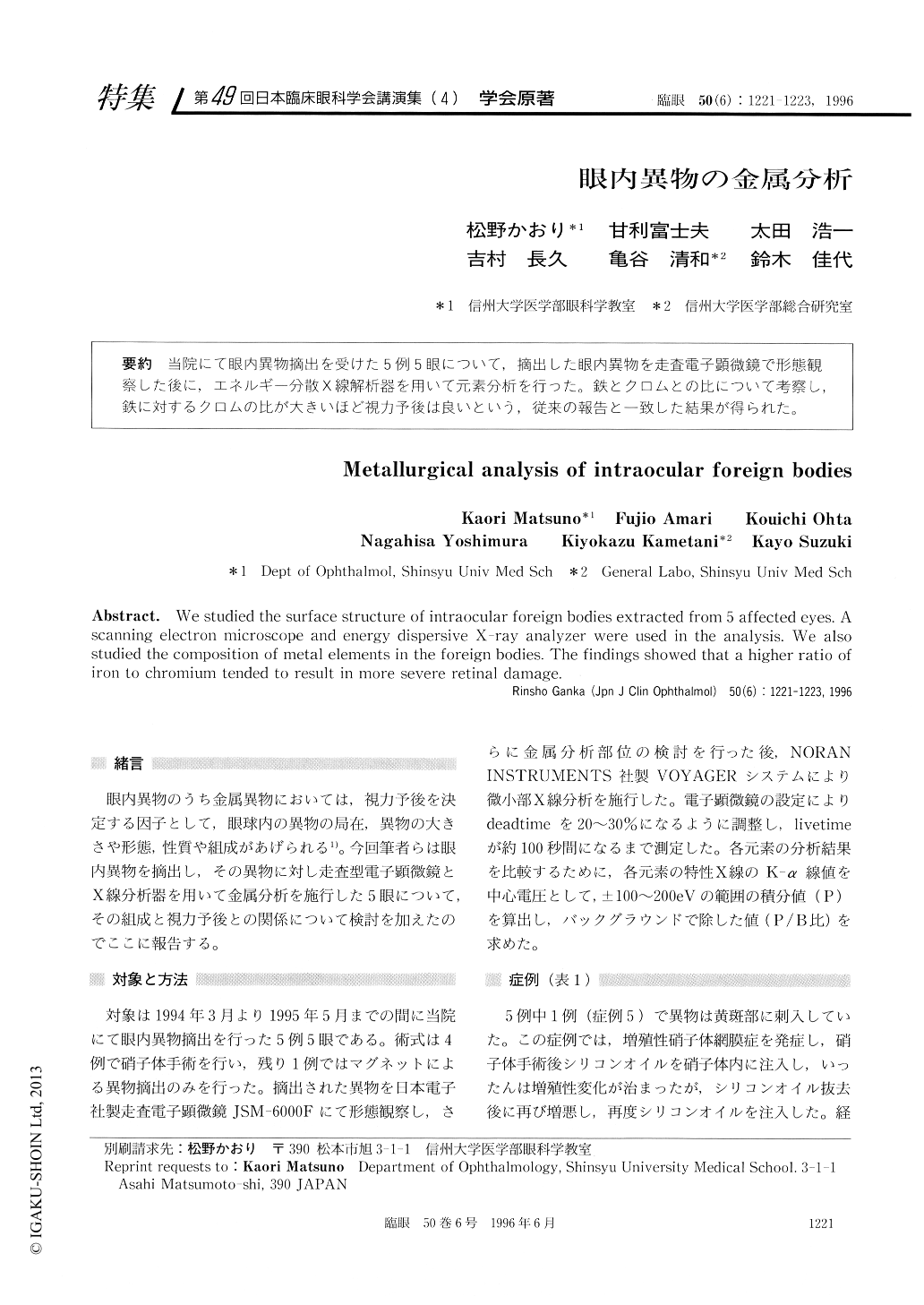
当院にて眼内異物摘出を受けた5例5眼について,摘出した眼内異物を走査電子顕微鏡で形態観察した後に,エネルギー分散X線解析器を用いて元素分析を行った。鉄とクロムとの比について考察し,鉄に対するクロムの比が大きいほど視力予後は良いという,従来の報告と一致した結果が得られた。
We studied the surface structure of intraocular foreign bodies extracted from 5 affected eyes. A scanning electron microscope and energy dispersive X-ray analyzer were used in the analysis. We also studied the composition of metal elements in the foreign bodies. The findings showed that a higher ratio of iron to chromium tended to result in more severe retinal damage.

Copyright © 1996, Igaku-Shoin Ltd. All rights reserved.


