Japanese
English
- 有料閲覧
PPV購入案内
1,320円 (1,200円+税10%) こちらは、695ページから698ページの文献です。購入には医書.jp本会員登録が必要です。
会員登録完了後に改めて上記「購入サイトへ」を選択してください。
または「購入サイトへ」選択後に「購入ログイン」、「新規会員登録」の順に進んでください。
会員登録について詳しくはをご確認ください。
- Abstract 文献概要
- 1ページ目 Look Inside
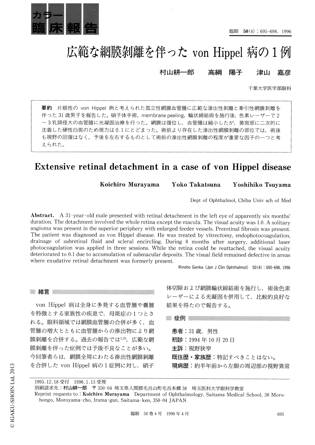
片眼性のvon Hippel病と考えられた孤立性網膜血管腫に広範な滲出性剥離と牽引性網膜剥離を伴った31歳男子を報告した。硝子体手術,membrane peeling,輪状締結術を施行後,色素レーザーで2〜3乳頭径大の血管腫に光凝固治療を行った。網膜は復位し,血管腫は縮小したが,黄斑部に二次的に沈着した硬性白斑のため視力は0.1にとどまった。術前より存在した滲出性網膜剥離の部位では,術後も視野の回復はなく,予後を左右するものとして術前の滲出性網膜剥離の程度が重要な因子の一つと考えられた。
A 31-year-old male presented with retinal detachment in the left eye of apparently six months' duration. The detachment involved the whole retina except the macula. The visual acuity was 1.0. A solitary angioma was present in the superior periphery with enlarged feeder vessels. Preretinal fibrosis was present. The patient was diagnosed as von Hippel disease. He was treated by vitrectomy, endophotocoagulation, drainage of subretinal fluid and scleral encircling. During 8 months after surgery, additional laser photocoagulation was applied in three sessions. While the retina could be reattached, the visual acuity deteriorated to 0.1 due to accumulation of submacular deposits. The visual field remained defective in areas where exudative retinal detachment was formerly present.
Copyright © 1996, Igaku-Shoin Ltd. All rights reserved.