Japanese
English
- 有料閲覧
PPV購入案内
1,320円 (1,200円+税10%) こちらは、35ページから39ページの文献です。購入には医書.jp本会員登録が必要です。
会員登録完了後に改めて上記「購入サイトへ」を選択してください。
または「購入サイトへ」選択後に「購入ログイン」、「新規会員登録」の順に進んでください。
会員登録について詳しくはをご確認ください。
- Abstract 文献概要
- 1ページ目 Look Inside
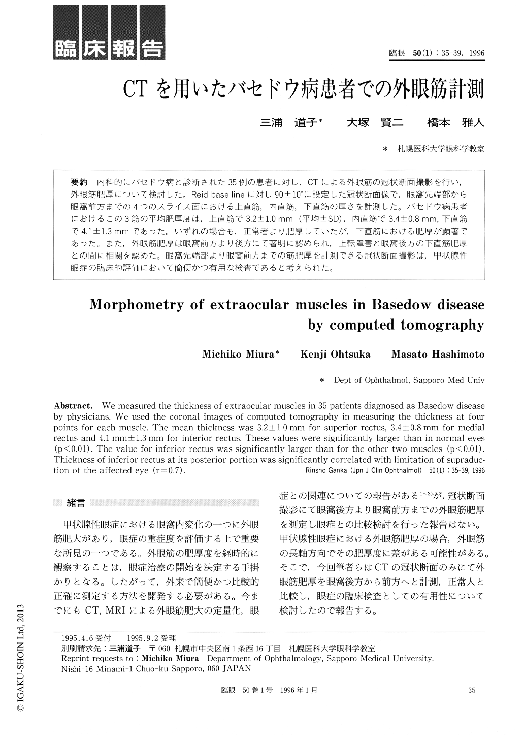
内科的にバセドウ病と診断された35例の患者に対し,CTによる外眼筋の冠状断面撮影を行い,外眼筋肥厚について検討した。Reid base lineに対し90±10°に設定した冠状断面像で,眼窩先端部から眼窩前方までの4つのスライス面における上直筋,内直筋,下直筋の厚さを計測した。バセドウ病患者におけるこの3筋の平均肥厚度は,上直筋で3.2±1.0mm (平均±SD),内直筋で3.4±0.8mm,下直筋で4.1±1.3mmであった。いずれの場合も,正常者より肥厚していたが,下直筋における肥厚が顕著であった。また,外眼筋肥厚は眼窩前方より後方にて著明に認められ,上転障害と眼窩後方の下直筋肥厚との間に相関を認めた。眼窩先端部より眼窩前方までの筋肥厚を計測できる冠状断面撮影は,甲状腺性眼症の臨床的評価において簡便かつ有用な検査であると考えられた。
We measured the thickness of extraocular muscles in 35 patients diagnosed as Basedow disease by physicians. We used the coronal images of computed tomography in measuring the thickness at four points for each muscle. The mean thickness was 3.2±1.0mm for superior rectus, 3.4±0.8mm for medial rectus and 4.1 mm±1.3mm for inferior rectus. These values were significantly larger than in normal eyes (p<0.01) . The value for inferior rectus was significantly larger than for the other two muscles (p<0.01).Thickness of inferior rectus at its posterior portion was significantly correlated with limitation of supraduc-tion of the affected eye (r=0.7).
Copyright © 1996, Igaku-Shoin Ltd. All rights reserved.