Japanese
English
- 有料閲覧
PPV購入案内
1,320円 (1,200円+税10%) こちらは、45ページから48ページの文献です。購入には医書.jp本会員登録が必要です。
会員登録完了後に改めて上記「購入サイトへ」を選択してください。
または「購入サイトへ」選択後に「購入ログイン」、「新規会員登録」の順に進んでください。
会員登録について詳しくはをご確認ください。
- Abstract 文献概要
- 1ページ目 Look Inside
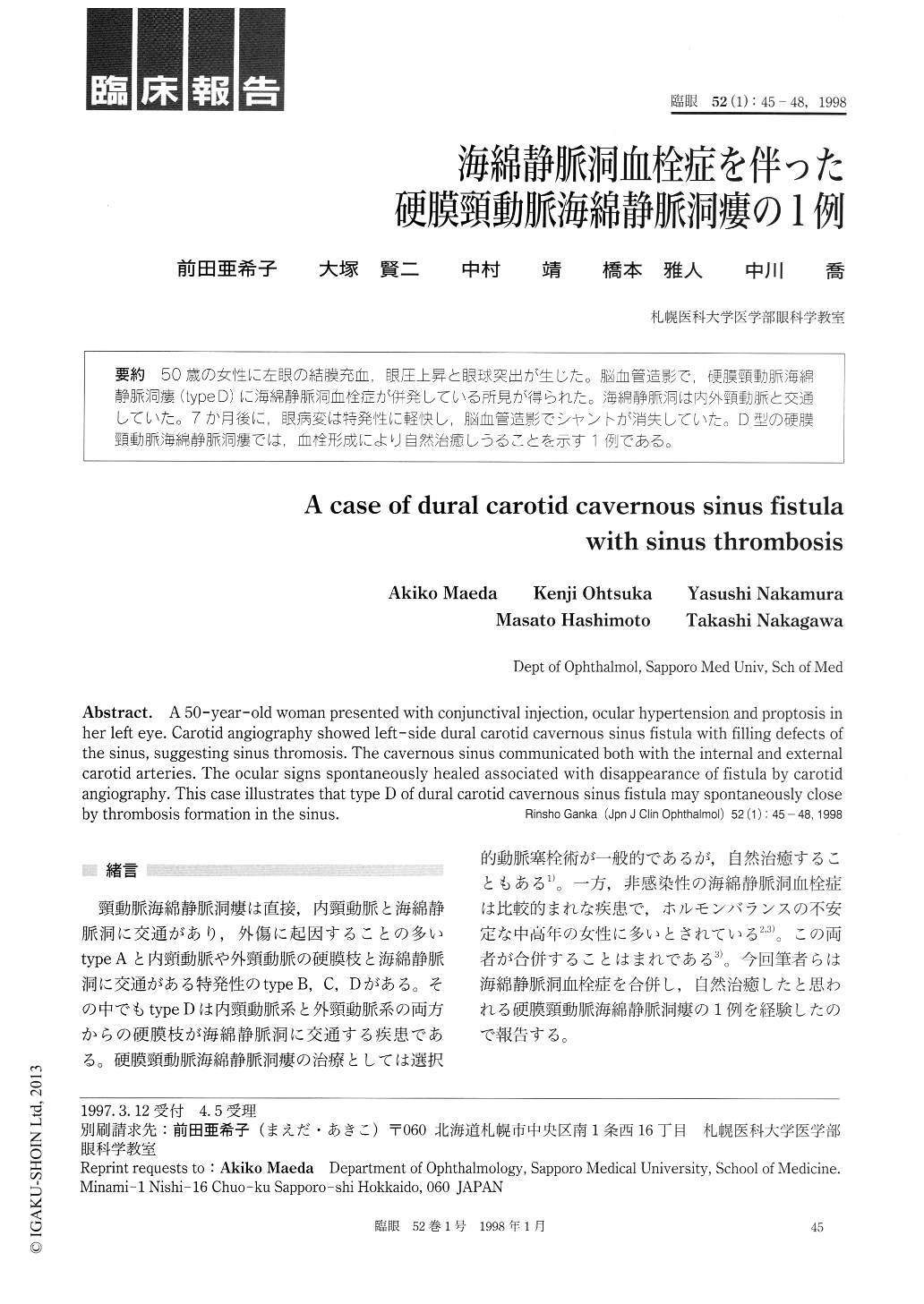
50歳の女性に左眼の結膜充血,眼圧上昇と眼球突出が生じた。脳血管造影で,硬膜頸動脈海綿静脈洞瘻(type D)に海綿静脈洞血栓症が併発している所見が得られた。海綿静脈洞は内外頸動脈と交通していた。7か月後に、眼病変は特発性に軽快し,脳血管造影でシャントが消失していた。D型の硬膜頸動脈海綿静脈洞瘻では、血栓形成により自然治癒しうることを示す1例である。
A 50-year-old woman presented with conjunctival injection, ocular hypertension and proptosis in her left eye. Carotid angiography showed left-side dural carotid cavernous sinus fistula with filling defects of the sinus, suggesting sinus thromosis. The cavernous sinus communicated both with the internal and external carotid arteries. The ocular signs spontaneously healed associated with disappearance of fistula by carotid angiography. This case illustrates that type D of dural carotid cavernous sinus fistula may spontaneously close by thrombosis formation in the sinus.
Copyright © 1998, Igaku-Shoin Ltd. All rights reserved.