Japanese
English
- 有料閲覧
PPV購入案内
1,320円 (1,200円+税10%) こちらは、1420ページから1424ページの文献です。購入には医書.jp本会員登録が必要です。
会員登録完了後に改めて上記「購入サイトへ」を選択してください。
または「購入サイトへ」選択後に「購入ログイン」、「新規会員登録」の順に進んでください。
会員登録について詳しくはをご確認ください。
- Abstract 文献概要
- 1ページ目 Look Inside
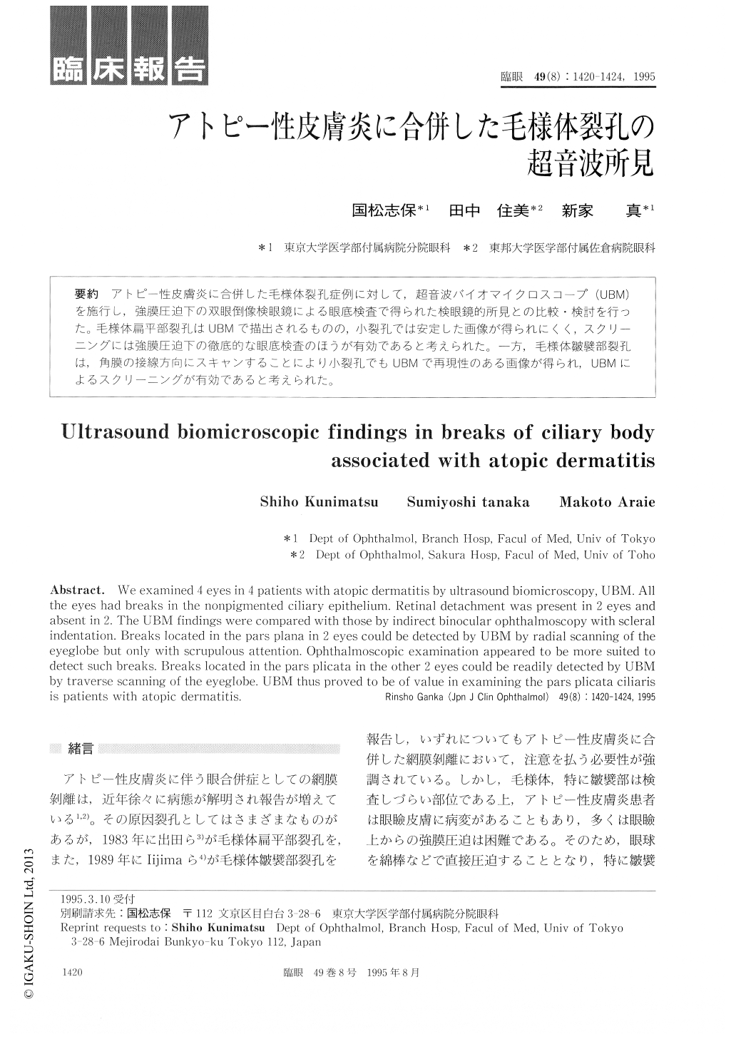
アトピー性皮膚炎に合併した毛様体裂孔症例に対して,超音波バイオマイクロスコープ(UBM)を施行し,強膜圧迫下の双眼倒像検眼鏡による眼底検査で得られた検眼鏡的所見との比較・検討を行った。毛様体扁平部裂孔はUBMで描出されるものの,小裂孔では安定した画像が得られにくく,スクリーニングには強膜圧迫下の徹底的な眼底検査のほうが有効であると考えられた。一方,毛様体皺襞部裂孔は,角膜の接線方向にスキャンすることにより小裂孔でもUBMで再現性のある画像が得られ,UBMによるスクリーニングが有効であると考えられた。
We examined 4 eyes in 4 patients with atopic dermatitis by ultrasound biomicroscopy, UBM. Allthe eyes had breaks in the nonpigmented ciliary epithelium. Retinal detachment was present in 2 eyes andabsent in 2. The UBM findings were compared with those by indirect binocular ophthalmoscopy with scleralindentation. Breaks located in the pars plana in 2 eyes could be detected by UBM by radial scanning of theeyeglobe but only with scrupulous attention. Ophthalmoscopic examination appeared to be more suited todetect such breaks. Breaks located in the pars plicata in the other 2 eyes. could be readily detected by UBMby traverse scanning of the eyeglobe. UBM thus proved to be of value in examining the pars plicata ciliarisis patients with atopic dermatitis.
Copyright © 1995, Igaku-Shoin Ltd. All rights reserved.