Japanese
English
- 有料閲覧
- Abstract 文献概要
- 1ページ目 Look Inside
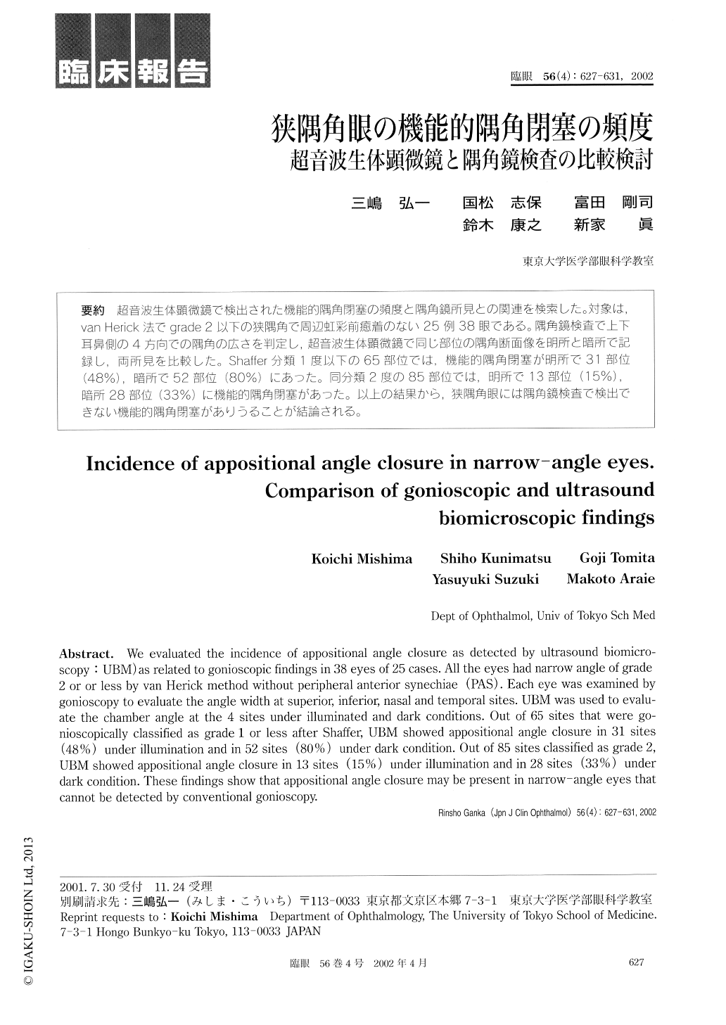
超音波生体顕微鏡で検出された機能的隅角閉塞の頻度と隅角鏡所見との関連を検索した。対象は,van Herick法でgrade 2以下の狭隅角で周辺虹彩前癒着のない25例38眼である。隅角鏡検査で上下耳鼻側の4方向での隅角の広さを判定し,超音波生体顕微鏡で同じ部位の隅角断面像を明所と暗所で記録し,両所見を比較した。Shaffer分類1度以下の65部位では,機能的隅角閉塞が明所で31部位(48%),暗所で52部位(80%)にあった。同分類2度の85部位では,明所で13部位(15%),暗所28部位(33%)に機能的隅角閉塞があった。以上の結果から,狭隅角眼には隅角鏡検査で検出できない機能的隅角閉塞がありうることが結論される。
We evaluated the incidence of appositional angle closure as detected by ultrasound biomicro-scopy :UBM) as related to gonioscopic findings in 38 eyes of 25 cases. All the eyes had narrow angle of grade 2 or or less by van Herick method without peripheral anterior synechiae (PAS). Each eye was examined by gonioscopy to evaluate the angle width at superior, inferior, nasal and temporal sites. UBM was used to evalu-ate the chamber angle at the 4 sites under illuminated and dark conditions. Out of 65 sites that were go-nioscopically classified as grade 1 or less after Shaffer, UBM showed appositional angle closure in 31 sites (48%) under illumination and in 52 sites (80%) under dark condition. Out of 85 sites classified as grade 2, UBM showed appositional angle closure in 13 sites (15%) under illumination and in 28 sites (33%) under dark condition. These findings show that appositional angle closure may be present in narrow-angle eyes that cannot be detected by conventional gonioscopy.

Copyright © 2002, Igaku-Shoin Ltd. All rights reserved.


