Japanese
English
- 有料閲覧
PPV購入案内
1,320円 (1,200円+税10%) こちらは、745ページから748ページの文献です。購入には医書.jp本会員登録が必要です。
会員登録完了後に改めて上記「購入サイトへ」を選択してください。
または「購入サイトへ」選択後に「購入ログイン」、「新規会員登録」の順に進んでください。
会員登録について詳しくはをご確認ください。
- Abstract 文献概要
- 1ページ目 Look Inside
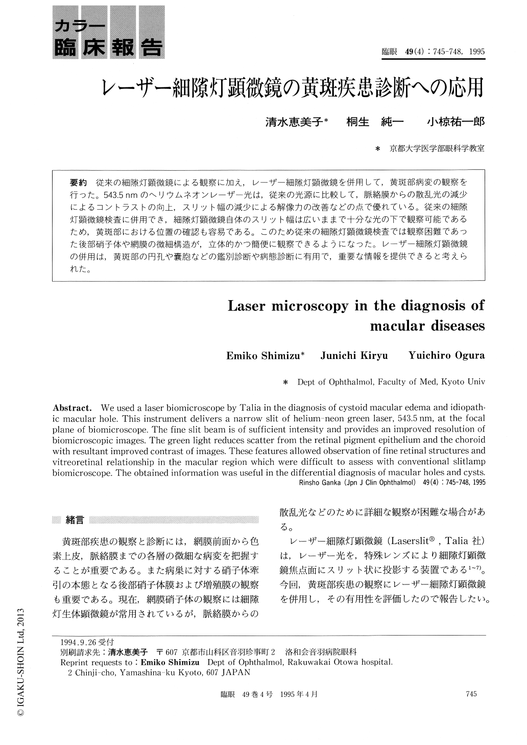
従来の細隙灯顕微鏡による観察に加え,レーザー細隙灯顕微鏡を併用して,黄斑部病変の観察を行った。543.5nmのヘリウムネオンレーザー光は,従来の光源に比較して,脈絡膜からの散乱光の減少によるコントラストの向上,スリット幅の減少による解像力の改善などの点で優れている。従来の細隙灯顕微鏡検査に併用でき,細隙灯顕微鏡自体のスリット幅は広いままで十分な光の下で観察可能であるため,黄斑部における位置の確認も容易である。このため従来の細隙灯顕微鏡検査では観察困難であった後部硝子体や網膜の微細構造が,立体的かつ簡便に観察できるようになった。レーザー細隙灯顕微鏡の併用は,黄斑部の円孔や嚢胞などの鑑別診断や病態診断に有用で,重要な情報を提供できると考えられた。
We used a laser biomicroscope by Talia in the diagnosis of cystoid macular edema and idiopath-ic macular hole. This instrument delivers a narrow slit of helium-neon green laser, 543.5nm, at the focal plane of biomicroscope. The fine slit beam is of sufficient intensity and provides an improved resolution of biomicroscopic images. The green light reduces scatter from the retinal pigment epithelium and the choroid with resultant improved contrast of images. These features allowed observation of fine retinal structures and vitreoretinal relationship in the macular region which were difficult to assess with conventional slitlamp biomicroscope. The obtained information was useful in the differential diagnosis of macular holes and cysts.
Copyright © 1995, Igaku-Shoin Ltd. All rights reserved.