Japanese
English
- 有料閲覧
PPV購入案内
1,320円 (1,200円+税10%) こちらは、759ページから762ページの文献です。購入には医書.jp本会員登録が必要です。
会員登録完了後に改めて上記「購入サイトへ」を選択してください。
または「購入サイトへ」選択後に「購入ログイン」、「新規会員登録」の順に進んでください。
会員登録について詳しくはをご確認ください。
- Abstract 文献概要
- 1ページ目 Look Inside
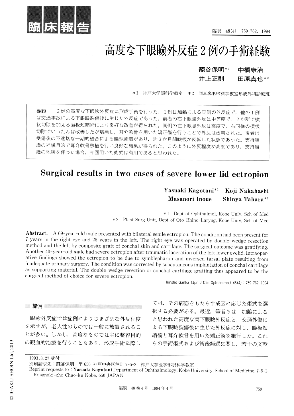
2例の高度な下眼瞼外反症に形成手術を行った。1例は加齢による両側の外反症で,他の1例は交通事故による下眼瞼裂傷後に生じた外反症であった。前者の右下眼瞼外反は中等度で,2か所で楔状切除を加える瞼板短縮術により良好な改善が得られた。同例の左下眼瞼外反は高度で,右同様の楔状切除でいったんは改善したが増悪し,耳介軟骨を用いた矯正術を行うことで外反は改善された。後者は受傷後の不適切な一期的縫合による瞼球癒着があり,約3か月間瞼板が反転した状態であった。支持組織の補填目的で耳介軟骨移植を行い良好な結果が得られた。このように外反程度が高度であり,支持組織の弛緩を伴った場合,今回用いた術式は有用であると思われた。
A 69-year-old male presented with bilateral senile ectropion. The condition had been present for 7 years in the right eye and 25 years in the left. The right eye was operated by double-wedge resection method and the left by composite graft of conchal skin and cartilage. The surgical outcome was gratifying. Another 40-year-old male had severe ectropion after traumatic laceration of the left lower eyelid. Intraoper-ative findings showed the ectropion to be due to symblepharon and inversed tarsal plate resulting from inadequate primary surgery. The condition was corrected by subcutaneous implantation of conchal cartilage as supporting material. The double-wedge resection or conchal cartilage grafting thus appeared to be the surgical method of choice for severe ectropion.
Copyright © 1994, Igaku-Shoin Ltd. All rights reserved.