Japanese
English
- 有料閲覧
PPV購入案内
1,320円 (1,200円+税10%) こちらは、1339ページから1341ページの文献です。購入には医書.jp本会員登録が必要です。
会員登録完了後に改めて上記「購入サイトへ」を選択してください。
または「購入サイトへ」選択後に「購入ログイン」、「新規会員登録」の順に進んでください。
会員登録について詳しくはをご確認ください。
- Abstract 文献概要
- 1ページ目 Look Inside
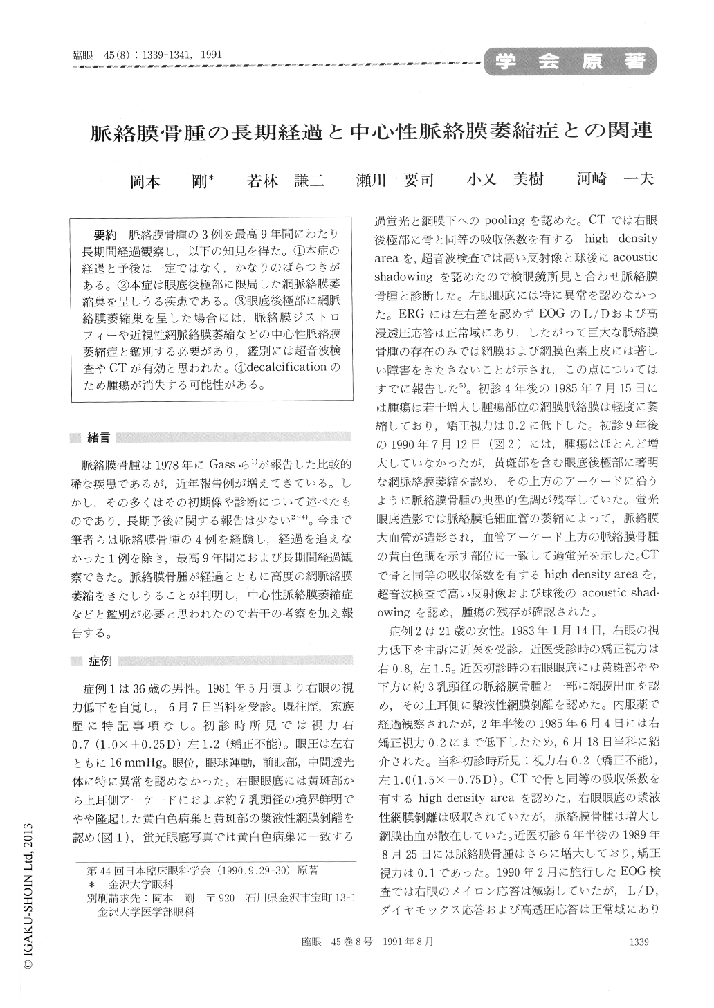
脈絡膜骨腫の3例を最高9年間にわたり長期間経過観察し,以下の知見を得た。①本症の経過と予後は一定ではなく,かなりのばらつきがある。②本症は眼底後極部に限局した網脈絡膜萎縮巣を呈しうる疾患である。③眼底後極部に網脈絡膜萎縮巣を呈した場合には,脈絡膜ジストロフィーや近視性網脈絡膜萎縮などの中心性脈絡膜萎縮症と鑑別する必要があり,鑑別には超音波検査やCTが有効と思われた。④decalcificationのため腫瘍が消失する可能性がある。
We followed up 3 cases of choroidal osteoma for up to 9 years. The size of the osteoma and course of visual acuity failed to show a definite pattern. The osteomatous lesion resulted in severe focal chorio-retinal atrophy in the posterior fundus. Ultrasono-graphy and computed tomography were useful indetecting the extent of osteoma. They were also of value in differentiating choroidal atrophy due to osteoma from atypical focal choroidal atrophy, high myopia and central choroidal dystrophy. Decalcification of choroidal osteoma occasionally resulted in regression of the tumor leaving an atypical choroidal atrophy.
Copyright © 1991, Igaku-Shoin Ltd. All rights reserved.