Japanese
English
- 有料閲覧
- Abstract 文献概要
- 1ページ目 Look Inside
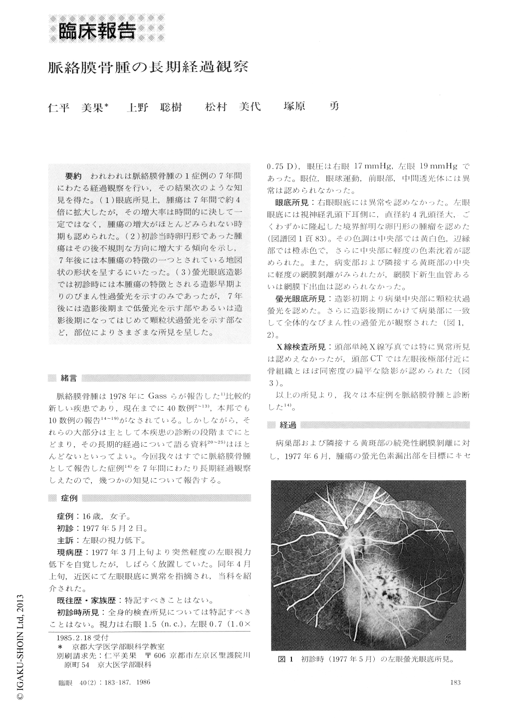
われわれは脈絡膜骨腫の1症列の7年間にわたる経過観察を行い,その結果次のような知見を得た.(1)眼底所見上,腫瘍は7年間で約4倍に拡大したが,その増大率は時間的に決して一定ではなく,腫瘍の増大がほとんどみられない時期も認められた.(2)初診当時卵円形であった腫瘍はその後不規則な方向に増大する傾向を示し,7年後には本腫瘍の特徴の一つとされている地図状の形状を呈するにいたった.(3)螢光眼底造影では初診時には本腫瘍の特徴とされる造影早期よりのびまん性過螢光を示すのみであったが,7年後には造影後期まで低螢光を示す部やあるいは造影後期になってはじめて顆粒状過螢光を示す部など,部位によりさまざまな所見を呈した.
We diagnosed unilateral osseous choristoma of the choroid in a 16-year-old girl and have followed up for 7 years. As the initial fundus manifestation, we observ-ed a yellowish-white and round subretinal tumor with sharply defined border. The tumor showed a slight elevation and was about 4 DD in diameter. Seven years later, the tumor attained a size of about 4 times in diameter. The initial round shape was transformed into a geographic pattern considered as typical for osseous choristoma. Fluorescein angiography showed char-acteristic hyperfluorescence of mottled pattern in the tumor area. The angiographic finding was not univorm nor constant, as some areas Showed hypofluorescence lasting till the after phase. Also, a granulated pattern was seen towards the late venous phase in the fundus area recently encroached by the tumor.
Rinsho Ganka (Jpn J Clin Ophthalmol) 40 (2) : 183-187, 1986

Copyright © 1986, Igaku-Shoin Ltd. All rights reserved.


