Japanese
English
- 有料閲覧
PPV購入案内
1,320円 (1,200円+税10%) こちらは、549ページから552ページの文献です。購入には医書.jp本会員登録が必要です。
会員登録完了後に改めて上記「購入サイトへ」を選択してください。
または「購入サイトへ」選択後に「購入ログイン」、「新規会員登録」の順に進んでください。
会員登録について詳しくはをご確認ください。
- Abstract 文献概要
- 1ページ目 Look Inside
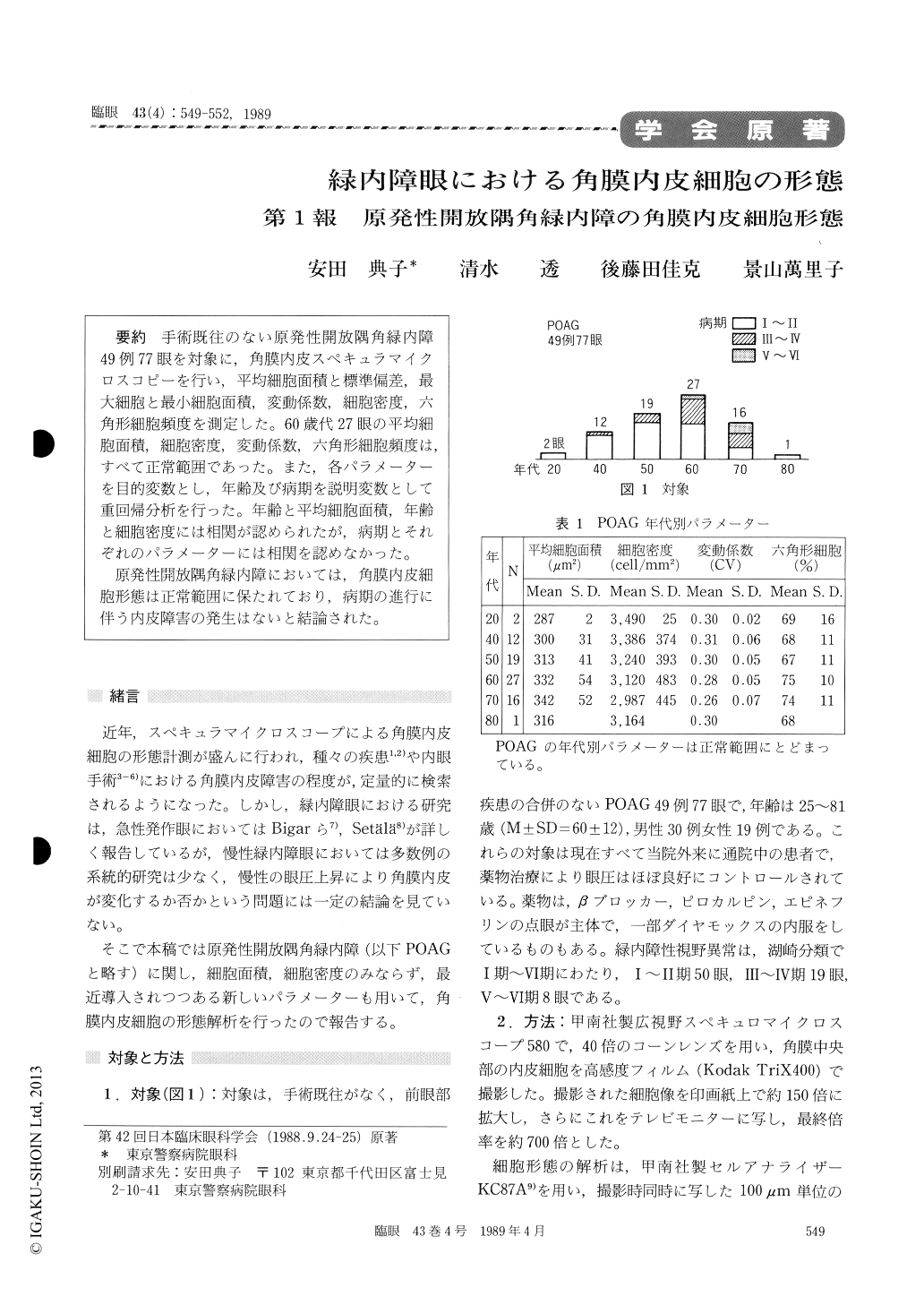
手術既往のない原発性開放隅角緑内障49例77眼を対象に,角膜内皮スペキュラマイクロスコピーを行い,平均細胞面積と標準偏差,最大細胞と最小細胞面積,変動係数,細胞密度,六角形細胞頻度を測定した。60歳代27眼の平均細胞面積,細胞密度,変動係数,六角形細胞頻度は,すべて正常範囲であった。また,各パラメーターを目的変数とし,年齢及び病期を説明変数として重回帰分析を行った。年齢と平均細胞面積,年齢と細胞密度には相関が認められたが,病期とそれぞれのパラメーターには相関を認めなかった。
原発性開放隅角緑内障においては,角膜内皮細胞形態は正常範囲に保たれており,病期の進行に伴う内皮障害の発生はないと結論された。
We performed specular microscopy in 77 eyes of 49 patients with primary open angle glaucoma. With the use of digitizer system, we computed the mean cell area, coefficient of variation, cell density and percentage of hexagonal cells in the corneal endothelium. In 27 eyes in 7th decade of age, these values were within normal range as compared with healthy controls.
We then performed a multivariate analysis by using the above parameters as criterion variables and by placing the age and the state of visual field as predictor variables. A positive correlation (p<O.05) was present between the age and mean cell area, and between the age and cell density. No correlation was present between the visual field and any of the parameters.
The findings suggest that the corneal endoth-elium is not noticeably affected in eyes with pri-mary open angle glaucoma.
Copyright © 1989, Igaku-Shoin Ltd. All rights reserved.