Japanese
English
- 有料閲覧
PPV購入案内
1,320円 (1,200円+税10%) こちらは、679ページから684ページの文献です。購入には医書.jp本会員登録が必要です。
会員登録完了後に改めて上記「購入サイトへ」を選択してください。
または「購入サイトへ」選択後に「購入ログイン」、「新規会員登録」の順に進んでください。
会員登録について詳しくはをご確認ください。
- Abstract 文献概要
- 1ページ目 Look Inside
- 参考文献 Reference
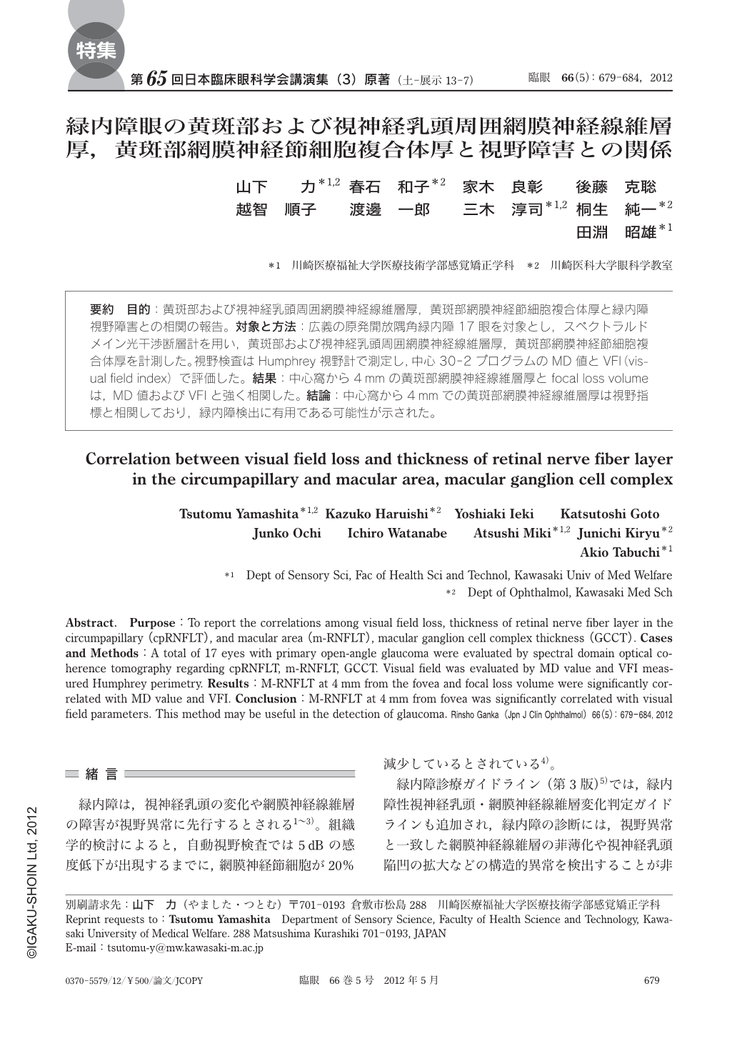
要約 目的:黄斑部および視神経乳頭周囲網膜神経線維層厚,黄斑部網膜神経節細胞複合体厚と緑内障視野障害との相関の報告。対象と方法:広義の原発開放隅角緑内障17眼を対象とし,スペクトラルドメイン光干渉断層計を用い,黄斑部および視神経乳頭周囲網膜神経線維層厚,黄斑部網膜神経節細胞複合体厚を計測した。視野検査はHumphrey視野計で測定し,中心30-2プログラムのMD値とVFI(visual field index)で評価した。結果:中心窩から4mmの黄斑部網膜神経線維層厚とfocal loss volumeは,MD値およびVFIと強く相関した。結論:中心窩から4mmでの黄斑部網膜神経線維層厚は視野指標と相関しており,緑内障検出に有用である可能性が示された。
Abstract. Purpose:To report the correlations among visual field loss,thickness of retinal nerve fiber layer in the circumpapillary(cpRNFLT),and macular area(m-RNFLT),macular ganglion cell complex thickness(GCCT). Cases and Methods:A total of 17 eyes with primary open-angle glaucoma were evaluated by spectral domain optical coherence tomography regarding cpRNFLT,m-RNFLT,GCCT. Visual field was evaluated by MD value and VFI measured Humphrey perimetry. Results:M-RNFLT at 4 mm from the fovea and focal loss volume were significantly correlated with MD value and VFI. Conclusion:M-RNFLT at 4 mm from fovea was significantly correlated with visual field parameters. This method may be useful in the detection of glaucoma.
Copyright © 2012, Igaku-Shoin Ltd. All rights reserved.