Japanese
English
- 有料閲覧
PPV購入案内
1,320円 (1,200円+税10%) こちらは、635ページから642ページの文献です。購入には医書.jp本会員登録が必要です。
会員登録完了後に改めて上記「購入サイトへ」を選択してください。
または「購入サイトへ」選択後に「購入ログイン」、「新規会員登録」の順に進んでください。
会員登録について詳しくはをご確認ください。
- Abstract 文献概要
- 1ページ目 Look Inside
- 参考文献 Reference
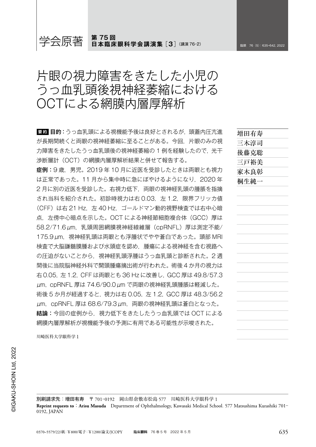
要約 目的:うっ血乳頭による視機能予後は良好とされるが,頭蓋内圧亢進が長期間続くと両眼の視神経萎縮に至ることがある。今回,片眼のみの視力障害をきたしたうっ血乳頭後の視神経萎縮の1例を経験したので,光干渉断層計(OCT)の網膜内層厚解析結果と併せて報告する。
症例:9歳,男児。2019年10月に近医を受診したときは両眼とも視力は正常であった。11月から集中時に急にぼやけるようになり,2020年2月に別の近医を受診した。右視力低下,両眼の視神経乳頭の腫脹を指摘され当科を紹介された。初診時視力は右0.03,左1.2,限界フリッカ値(CFF)は右21Hz,左40Hz,ゴールドマン動的視野検査では右中心暗点,左傍中心暗点を示した。OCTによる神経節細胞複合体(GCC)厚は58.2/71.6μm,乳頭周囲網膜視神経線維層(cpRNFL)厚は測定不能/175.9μm,視神経乳頭は両眼とも浮腫状でやや蒼白であった。頭部MRI検査で大脳鎌髄膜腫および水頭症を認め,腫瘍による視神経を含む視路への圧迫がないことから,視神経乳頭浮腫はうっ血乳頭と診断された。2週間後に当院脳神経外科で開頭腫瘍摘出術が行われた。術後4か月の視力は右0.05,左1.2,CFFは両眼とも36Hzに改善し,GCC厚は49.8/57.3μm,cpRNFL厚は74.6/90.0μmで両眼の視神経乳頭腫脹は軽減した。術後5か月が経過すると,視力は右0.05,左1.2,GCC厚は48.3/56.2μm,cpRNFL厚は68.6/79.3μm,両眼の視神経乳頭は蒼白となった。
結論:今回の症例から,視力低下をきたしたうっ血乳頭ではOCTによる網膜内層厚解析が視機能予後の予測に有用である可能性が示唆された。
Abstract Purpose:The prognosis for visual function due to congestion of papillae is good, but prolonged increased intracranial pressure can lead to optic atrophy in both eyes. We report a case of optic atrophy after the papilledema that resulted in vision loss in only one eye.
Case:The case was a 9-year-old boy. Visual acuity was normal in both eyes when he had visited a nearby ophthalmologist in October 2019. In November, he suddenly began to blur when concentrating, and in February 2020, he visited a local ophthalmologist. The patient was referred to our department because of decreased visual acuity in the right eye and optic disc edema in both eyes. Visual acuity at first visit was 0.03 right, 1.2 left, 21 Hz right, and 40 Hz left with critical flicker frequency(CFF). OCT showed GCC thickness of 58.2/71.6 μm, cpRNFL thickness of unmeasurable/175.9 μm, and optic disc was edematous and slightly pale in both eyes. Head MRI showed falx meningioma, and optic disc edema was diagnosed as papilledema because the tumor did not compress the optic tract, including the optic nerve. Two weeks later, a craniotomy tumor was removed by neurosurgery. Four months after surgery, visual acuity improved to 0.05 right and 1.2 left;CFF improved to 36 Hz in both eyes, GCC thickness was 49.8/57.3 μm, cpRNFL thickness was 74.6/90.0 μm, and optic disc swelling was reduced in both eyes. After 5 months from the operation, visual acuity was 0.05 right and 1.2 left, GCC thickness was 48.3/56.2 μm, cpRNFL thickness was 68.6/79.3 μm, and optic disc became pale in both eyes.
Conclusion:Prognosis of optic atrophy after the papilledema might be different in this case by OCT.
Copyright © 2022, Igaku-Shoin Ltd. All rights reserved.