Japanese
English
- 有料閲覧
PPV購入案内
1,320円 (1,200円+税10%) こちらは、1371ページから1375ページの文献です。購入には医書.jp本会員登録が必要です。
会員登録完了後に改めて上記「購入サイトへ」を選択してください。
または「購入サイトへ」選択後に「購入ログイン」、「新規会員登録」の順に進んでください。
会員登録について詳しくはをご確認ください。
- Abstract 文献概要
- 1ページ目 Look Inside
- 参考文献 Reference
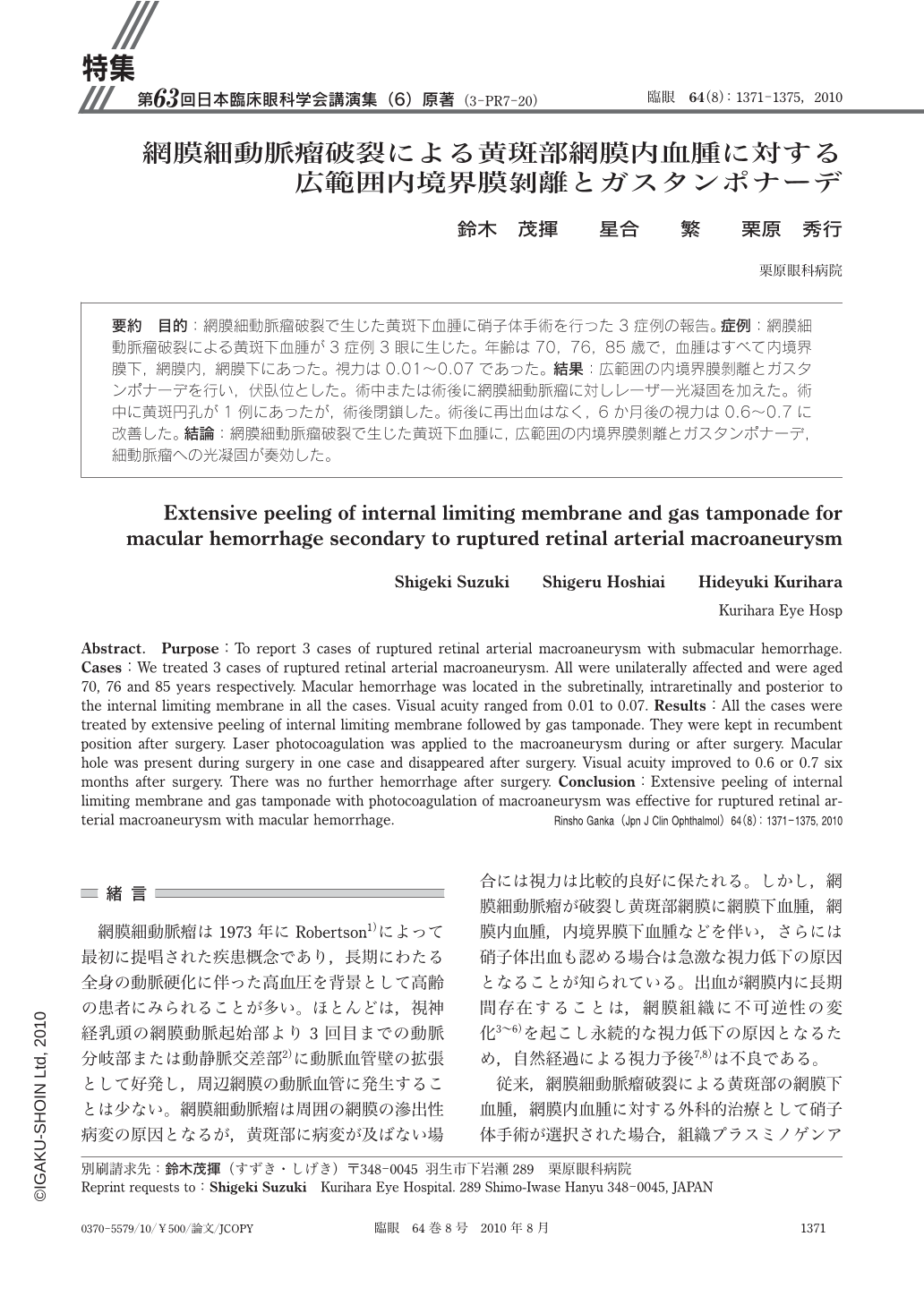
要約 目的:網膜細動脈瘤破裂で生じた黄斑下血腫に硝子体手術を行った3症例の報告。症例:網膜細動脈瘤破裂による黄斑下血腫が3症例3眼に生じた。年齢は70,76,85歳で,血腫はすべて内境界膜下,網膜内,網膜下にあった。視力は0.01~0.07であった。結果:広範囲の内境界膜剝離とガスタンポナーデを行い,伏臥位とした。術中または術後に網膜細動脈瘤に対しレーザー光凝固を加えた。術中に黄斑円孔が1例にあったが,術後閉鎖した。術後に再出血はなく,6か月後の視力は0.6~0.7に改善した。結論:網膜細動脈瘤破裂で生じた黄斑下血腫に,広範囲の内境界膜剝離とガスタンポナーデ,細動脈瘤への光凝固が奏効した。
Abstract. Purpose:To report 3 cases of ruptured retinal arterial macroaneurysm with submacular hemorrhage. Cases:We treated 3 cases of ruptured retinal arterial macroaneurysm. All were unilaterally affected and were aged 70,76 and 85 years respectively. Macular hemorrhage was located in the subretinally,intraretinally and posterior to the internal limiting membrane in all the cases. Visual acuity ranged from 0.01 to 0.07. Results:All the cases were treated by extensive peeling of internal limiting membrane followed by gas tamponade. They were kept in recumbent position after surgery. Laser photocoagulation was applied to the macroaneurysm during or after surgery. Macular hole was present during surgery in one case and disappeared after surgery. Visual acuity improved to 0.6 or 0.7 six months after surgery. There was no further hemorrhage after surgery. Conclusion:Extensive peeling of internal limiting membrane and gas tamponade with photocoagulation of macroaneurysm was effective for ruptured retinal arterial macroaneurysm with macular hemorrhage.
Copyright © 2010, Igaku-Shoin Ltd. All rights reserved.