Japanese
English
- 有料閲覧
PPV購入案内
1,320円 (1,200円+税10%) こちらは、551ページから554ページの文献です。購入には医書.jp本会員登録が必要です。
会員登録完了後に改めて上記「購入サイトへ」を選択してください。
または「購入サイトへ」選択後に「購入ログイン」、「新規会員登録」の順に進んでください。
会員登録について詳しくはをご確認ください。
- Abstract 文献概要
- 1ページ目 Look Inside
- 参考文献 Reference
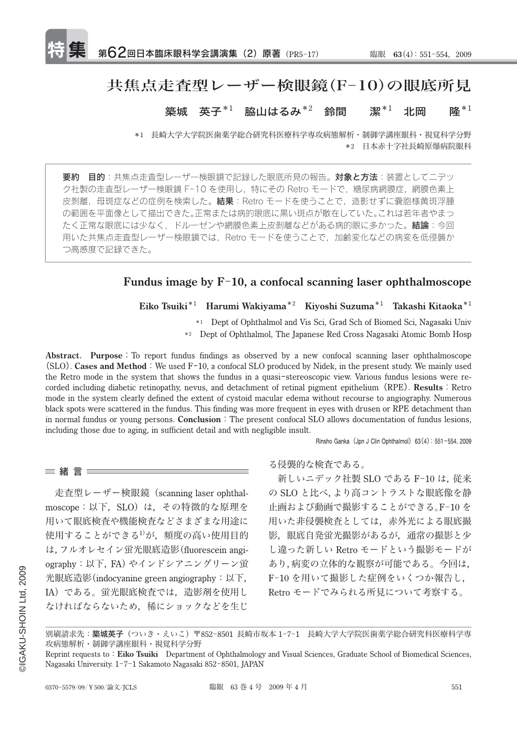
要約 目的:共焦点走査型レーザー検眼鏡で記録した眼底所見の報告。対象と方法:装置としてニデック社製の走査型レーザー検眼鏡F-10を使用し,特にそのRetroモードで,糖尿病網膜症,網膜色素上皮剝離,母斑症などの症例を検索した。結果:Retroモードを使うことで,造影せずに囊胞様黄斑浮腫の範囲を平面像として描出できた。正常または病的眼底に黒い斑点が散在していた。これは若年者やまったく正常な眼底には少なく,ドルーゼンや網膜色素上皮剝離などがある病的眼に多かった。結論:今回用いた共焦点走査型レーザー検眼鏡では,Retroモードを使うことで,加齢変化などの病変を低侵襲かつ高感度で記録できた。
Abstract. Purpose:To report fundus findings as observed by a new confocal scanning laser ophthalmoscope(SLO). Cases and Method:We used F-10,a confocal SLO produced by Nidek,in the present study. We mainly used the Retro mode in the system that shows the fundus in a quasi-stereoscopic view. Various fundus lesions were recorded including diabetic retinopathy,nevus,and detachment of retinal pigment epithelium(RPE). Results:Retro mode in the system clearly defined the extent of cystoid macular edema without recourse to angiography. Numerous black spots were scattered in the fundus. This finding was more frequent in eyes with drusen or RPE detachment than in normal fundus or young persons. Conclusion:The present confocal SLO allows documentation of fundus lesions,including those due to aging,in sufficient detail and with negligible insult.
Copyright © 2009, Igaku-Shoin Ltd. All rights reserved.