Japanese
English
- 有料閲覧
PPV購入案内
1,320円 (1,200円+税10%) こちらは、841ページから844ページの文献です。購入には医書.jp本会員登録が必要です。
会員登録完了後に改めて上記「購入サイトへ」を選択してください。
または「購入サイトへ」選択後に「購入ログイン」、「新規会員登録」の順に進んでください。
会員登録について詳しくはをご確認ください。
- Abstract 文献概要
- 1ページ目 Look Inside
- 参考文献 Reference
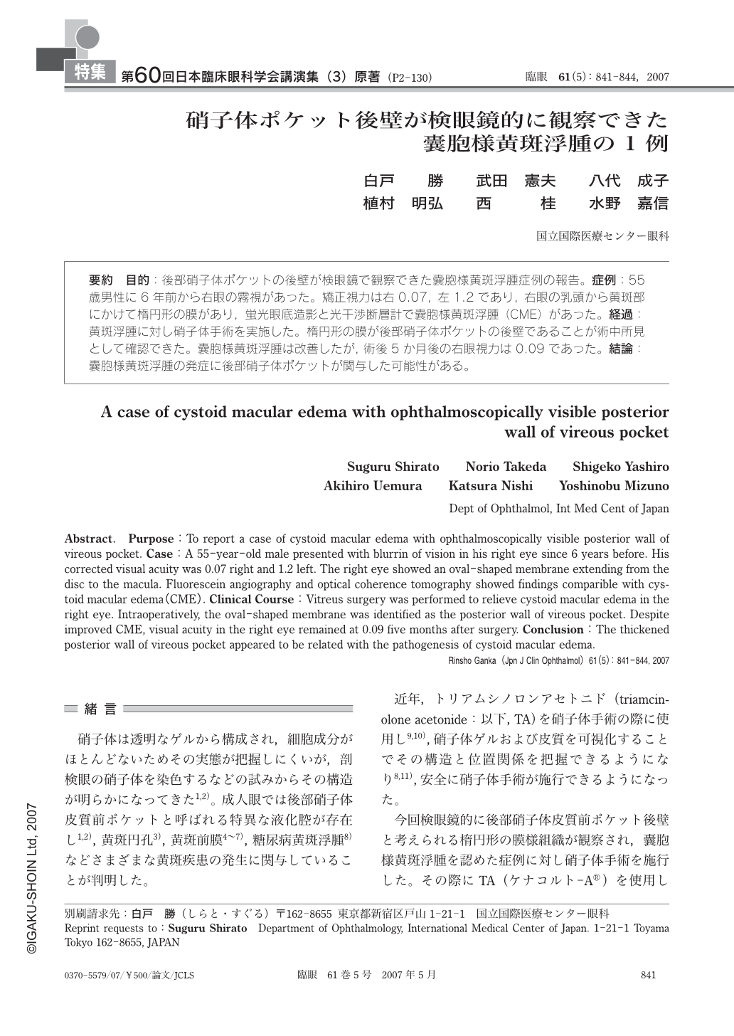
要約 目的:後部硝子体ポケットの後壁が検眼鏡で観察できた囊胞様黄斑浮腫症例の報告。症例:55歳男性に6年前から右眼の霧視があった。矯正視力は右0.07,左1.2であり,右眼の乳頭から黄斑部にかけて楕円形の膜があり,蛍光眼底造影と光干渉断層計で囊胞様黄斑浮腫(CME)があった。経過:黄斑浮腫に対し硝子体手術を実施した。楕円形の膜が後部硝子体ポケットの後壁であることが術中所見として確認できた。囊胞様黄斑浮腫は改善したが,術後5か月後の右眼視力は0.09であった。結論:囊胞様黄斑浮腫の発症に後部硝子体ポケットが関与した可能性がある。
Abstract. Purpose:To report a case of cystoid macular edema with ophthalmoscopically visible posterior wall of vireous pocket. Case:A 55-year-old male presented with blurrin of vision in his right eye since 6 years before. His corrected visual acuity was 0.07 right and 1.2 left. The right eye showed an oval-shaped membrane extending from the disc to the macula. Fluorescein angiography and optical coherence tomography showed findings comparible with cystoid macular edema(CME). Clinical Course:Vitreus surgery was performed to relieve cystoid macular edema in the right eye. Intraoperatively, the oval-shaped membrane was identified as the posterior wall of vireous pocket. Despite improved CME, visual acuity in the right eye remained at 0.09 five months after surgery. Conclusion:The thickened posterior wall of vireous pocket appeared to be related with the pathogenesis of cystoid macular edema.
Copyright © 2007, Igaku-Shoin Ltd. All rights reserved.