Japanese
English
- 有料閲覧
PPV購入案内
1,320円 (1,200円+税10%) こちらは、1259ページから1262ページの文献です。購入には医書.jp本会員登録が必要です。
会員登録完了後に改めて上記「購入サイトへ」を選択してください。
または「購入サイトへ」選択後に「購入ログイン」、「新規会員登録」の順に進んでください。
会員登録について詳しくはをご確認ください。
- Abstract 文献概要
- 1ページ目 Look Inside
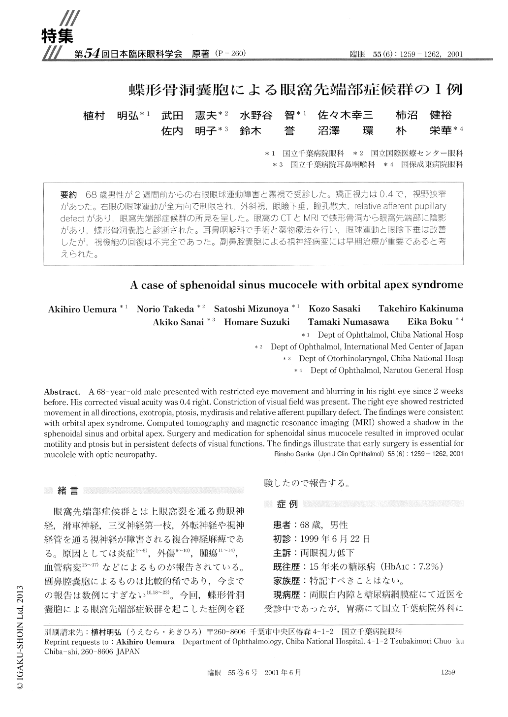
68歳男性が2週間前からの右眼眼球運動障害と霧視で受診した。矯正視力は0.4で,視野狭窄があった。右眼の眼球運動が全方向で制限され,外斜視,眼瞼下垂,瞳孔散大,relative afferent pupillarydefectがあり,眼窩先端部症候群の所見を呈した。眼窩のCTとMRIで蝶形骨洞から眼窩先端部に陰影があり,蝶形骨洞嚢胞と診断された。耳鼻咽喉科で手術と薬物療法を行い,眼球運動と眼瞼下垂は改善したが,視機能の回復は不完全であった。副鼻腔嚢胞による視神経病変には早期治療が重要であると考えられた。
A 68-year-old male presented with restricted eye movement and blurring in his right eye since 2 weeks before. His corrected visual acuity was 0.4 right. Constriction of visual field was present. The right eye showed restricted movement in all directions, exotropia, ptosis, mydirasis and relative afferent pupillary defect. The findings were consistent with orbital apex syndrome. Computed tomography and magnetic resonance imaging (MRI) showed a shadow in the sphenoidal sinus and orbital apex. Surgery and medication for sphenoidal sinus mucocele resulted in improved ocular motility and ptosis but in persistent defects of visual functions. The findings illustrate that early surgery is essential for mucolele with optic neuropathy.
Copyright © 2001, Igaku-Shoin Ltd. All rights reserved.